Japanese
English
- 有料閲覧
- Abstract 文献概要
- 1ページ目 Look Inside
術前診断が可能であったⅡb型早期胃癌の報告はきわめてまれである.さらにまた最大径が5mm以下の癌は微小癌として特別に扱われる傾向にあるが,微小癌の中でも典型Ⅱbは特殊な例を除いて臨床診断の対象にはならないとさえいわれている.著者らは臨床的に診断可能であり,切除胃の組織学的検討から確かめられた5mm以下の微小典型Ⅱb型早期胃癌を経験したので報告する.
A 60-year-old woman periodically visited the outpatient clinic in our hospital since one year and six months before the establishment of the present diag nosis. In April 1972 endoscopy of the stomach revealed a map-like reddened area in the upper part of the body on the posterior wall near the lesser curvature. Its oral side was seen to adjoin the transitional zone between the stomach esophagus, while its anterior wall side was not so clear-cut. On the posterior wall side the reddening was prominent in parts, to be distinguished in its margins from the surrounding mucosa. The reddened area contained bleeding spots. Biopsy from this area was twice attempted: cancer was positive in one of seven pieces in the first biopsy and in two of seven in the second. Biopsy specimens from the mid-body and from around the cardia were all negative for malignancy.
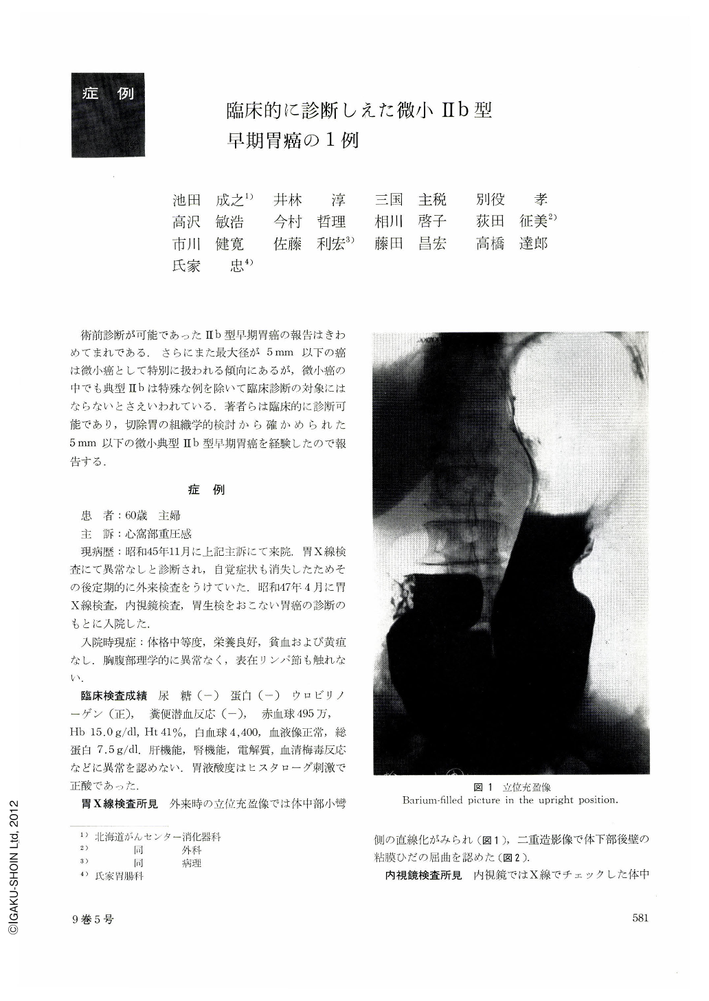
In radiography several very small radiolucencies were noticed in double contrast view almost in the same place as the presumed lesion on the posterior wall of the upper body. As compared with the normal areae gastricae, they were rather rougher, larger and more roundish.
Gross examination of both the fixed and unfixed specimens completely failed to locate the site of cancer. Histologically, it was adenocarcinoma tubulare, locatized within the mucosal layer and about 3×5 mm in size, located in the upper part of the body on the posterior wall near the lesser curvature. A strip of Ul-IIS about 10 mm long was seen on the lesser curvature adjacent to the cancer lesion.
Abnormal findings seen in both x-ray and endoscopy are believed to represent these mucosal changes including the minute cancer.

Copyright © 1974, Igaku-Shoin Ltd. All rights reserved.


