Japanese
English
- 有料閲覧
- Abstract 文献概要
- 1ページ目 Look Inside
症例
患者;64歳 主婦
主訴:心窩部重圧感
家族歴・既応歴:特記すべきことなし
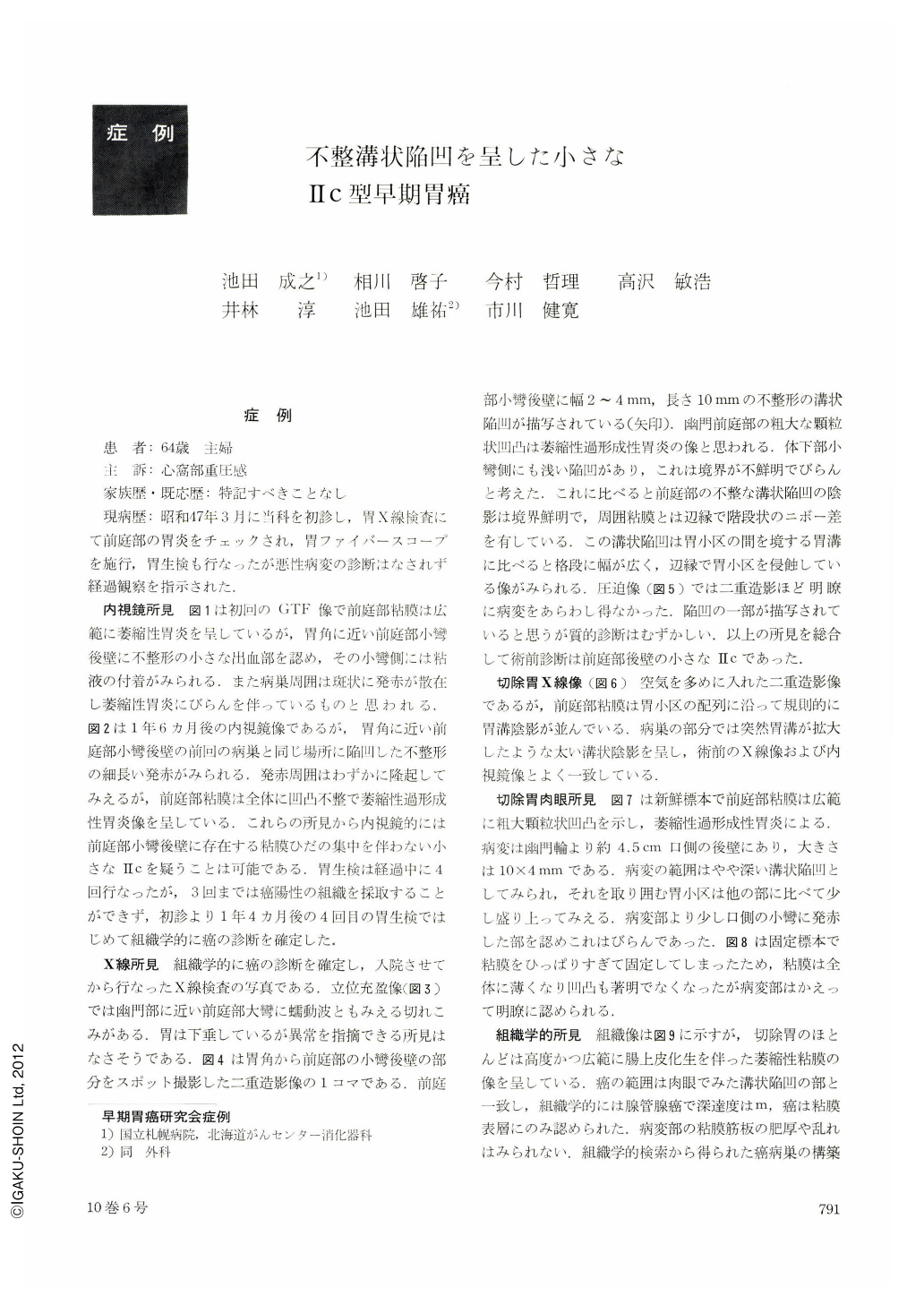
現病歴:昭和47年3月に当科を初診し,胃X線険査にて前庭部の胃炎をチェックされ,胃ファイバースコープを施行,胃生検も行なったが悪性病変の診断はなされず経過観察を指示された.
The patient: a 64-year-old woman. The initial GTF picture showed a small bleeding area of irregular shape on the posterior wall of the antrum near the lesser curvature of the angle. The antral mucosa presented a picture of extensive atrophic gastritis. Endoscopy one and a half year later revealed no bleeding area. Only a long strip of depression was revealed instead. The whole course of time up to the correct diagnosis of cancer was one year and four months. In the interim gastric biopsy was performed four times, but it invariably turned out negative for cancer until the fourth examination because we were unable to obtain appropriate specimens from the lesion up to then. In x-ray a groove-like depression of irregular shape, 2~4 mm wide and 10 mm long, was depicted on the posterior wall of the antrum near the lesser curvature. This groove-like depression was far wider than the sulci between the areae gastricae, eroding the areae in its margins. Resected stomach showed a sulcus-like depression on the posterior wall of the antrum 4.5 cm oral from the pyloric ring. The gastric areas surrounding it were slightly elevated as compared with other regions. Histologically it was tubular adenocarcinoma. Almost all the parts of the resected stomach displayed atrophic mucosa associated with advanced and extensive intestinal metaplasia.

Copyright © 1975, Igaku-Shoin Ltd. All rights reserved.


