Japanese
English
- 有料閲覧
- Abstract 文献概要
- 1ページ目 Look Inside
膵疾患の診断に関して血管造影の提供する情報はきわめて多大であり,最近では各施設においても経験の積み重ねが行なわれ,膵の血管造影は,膵癌をはじめとする各種膵疾患診断の必須検査となってきた.今回は1974年3月までに教室で経験した129例の膵癌に対して施行された動脈造影84例,静脈造影77例について検討を加え,筆者らが行なっている血管計測処理による膵癌診断への新しい試みについて述べる.また血管造影と各種検査法との併用診断および読影上鑑別のきわめて困難な慢性膵炎との相違についても自験例から検討を加えてみたい.
膵癌における血管造影の占める位置
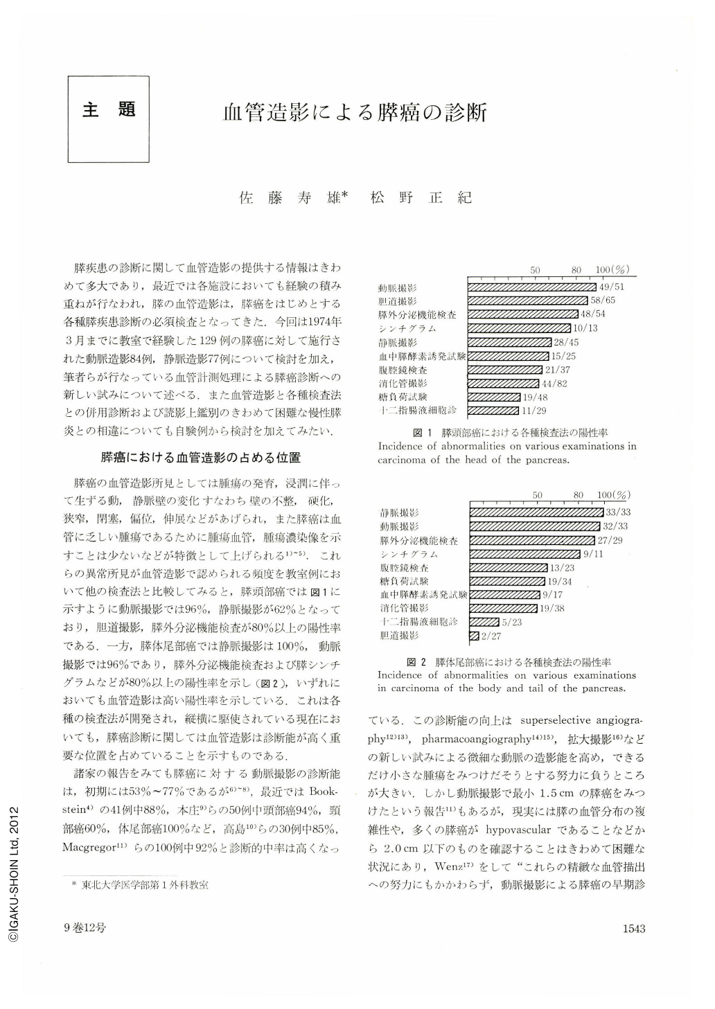
膵癌の血管造影所見としては腫瘍の発育,浸潤に伴って生ずる動,静脈壁の変化すなわち壁の不整,硬化,狭窄,閉塞,偏位,伸展などがあげられ,また膵癌は血管に乏しい腫瘍であるために腫瘍血管,腫瘍濃染像を示すことは少ないなどが特徴として上げられる1)~5).これらの異常所見が血管造影で認められる頻度を教室例において他の検査法と比較してみると,膵頭部癌では図1に示すように動脈撮影では96%,静脈撮影が62%となっており,胆道撮影,膵外分泌機能検査が80%以上の陽性率である.一方,膵体尾部癌では静脈撮影は100%,動脈撮影では96%であり,膵外分泌機能検査および膵シンチグラムなどが80%以上の陽性率を示し(図2),いずれにおいても血管造影は高い陽性率を示している.これは各種の検査法が開発され,縦横に駆使されている現在においても,膵癌診断に関しては血管造影は診断能が高く重要な位置を占めていることを示すものである.
We have performed selective angiography in 84 cases of pancreatic carcinoma and splenoportography in another 77 cases of it. Positive rate of abnormal findings in carcinoma of the head of the pancreas was 96 per cent in angiography and 62 per cent in portography, while in carcinoma of the body and tail of the pancreas it was 96 per cent in angiography and 100 per cent in portography. Despite such a high degree of accurate diagnosis the rate of resectability remained as low as 24 per cent in carcinoma of the pancreatic head and 17 per cent in that of the body and tail. Accurate diagnosis at an earlier poriod is thus highly desirable. It is emphasized here that in order to achieve yet higher rate of correct diagnosis in carcinoma of the pancreas more comprehensible judgment combined with other diagnostic techniques is essential.
For the purpose of grasping more minute positive findings, we have also calculated the estimated blood flow by measuring the size of the various arteries and splenic vein on the films of angiography and portography and obtained the following results.
1. The estimated blood flow from the celiac artery to the pancreas was in normal cases on an average 74.1 ml/min. In chronic pancreatitis the blood flow decreased, but it remained the same in cancer cases.
2. Distribution of the blood flow in the area of the head and that of the tail and body showed that in carcinoma the blood flow decreased in the affected area, resulting in its increase in the other area.
3. When the radius of the takeoffs in anterior, posterior superior pancreaticoduodenal arteries and in dorsal pancreatic artery were more than 0.75 mm wide, most of the third branches were visualized.
4. The estimated blood flow of the area that showed shadow defect in scanning was found to be decreased.
5. As compared with normal cases, those of carcinoma of the pancreatic head showed narrowing in the diameter of the splenic vein in the tail, and vein the was elongated.

Copyright © 1974, Igaku-Shoin Ltd. All rights reserved.


