Japanese
English
- 有料閲覧
- Abstract 文献概要
- 1ページ目 Look Inside
- サイト内被引用 Cited by
はじめに
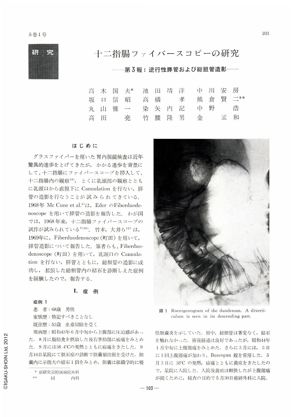
グラスファイバーを用いた胃内視鏡検査は近年驚異的進歩をとげてきたが,かかる進歩を背景にして,十二指腸にファイバースコープを挿入して,十二指腸内の観察13),とくに乳頭部の観察とともに乳頭口から直視下にCannulationを行ない,膵管の造影を行なうことが試みられてきている.1968年Mc Cune et al2)は,EderのFiberduodenoscopeを用いて膵管の造影を報告した.わが国では,1968年来,十二指腸ファイバースコープの試作が試みられている7)10).竹本,大井ら12)は,1969年に,Fiberduodenoscope(町田)を用いて,膵管造影について報告した.筆者らも,Fiberduodenoscope(町田)を用いて,乳頭口のCannulationを行ない,膵管とともに,総胆管の造影に成功し,拡張した総胆管内の結石を診断しえた症例を経験したので,報告する.
The common bile duct as well as the pancreatic one has been visualized by cannulation under direct vision into the ampulla of Vater by means of a fiberduodenoscope manufactured for trial (Machida) in a 60-year-old male and 71-year-old male. A shadow defect due to a calculus was demonstrated in the dilated common bile duct in both cases. Choledocholithiasis was later confirmed at operation. The serum amylase increased abptly but returned to the normal level within 1 week in the subjects whose whole pancreas was visualized radiologically, especially after pancreatography.
Choledochography and pancreatography by means of endoscopic cannulation the ampulla of Vater are also discussed correlating its changes caused by choledocholithiasis with the opening form of the common bile duct and the pancreatic one in the ampulla.

Copyright © 1970, Igaku-Shoin Ltd. All rights reserved.


