Japanese
English
- 有料閲覧
- Abstract 文献概要
- 1ページ目 Look Inside
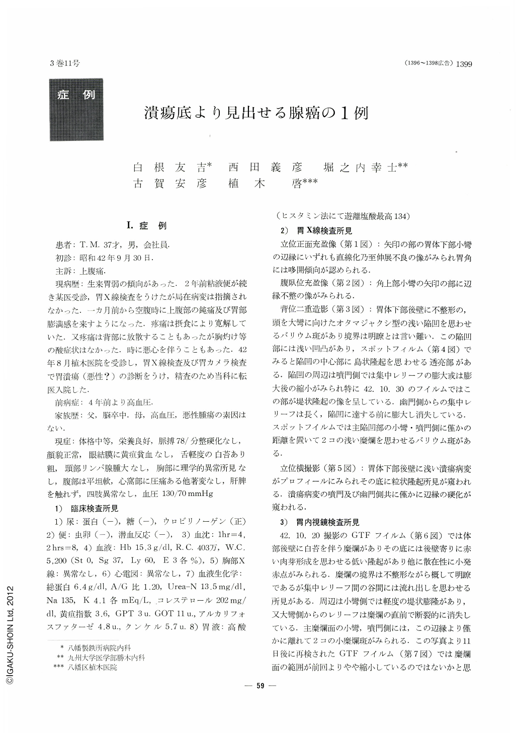
Ⅰ.症例
患者:T. M. 37才,男,会社員.
初診:昭和42年9月30日,
主訴:上腹痛.
現病歴:生来胃弱の傾向があった.2年前粘液便が続き某医受診,胃X線検査をうけたが局在病変は指摘されなかった.一カ月前から空腹時に上腹部の鈍痛及び胃部膨満感を来すようになった.疼痛は摂食により寛解していた.又疼痛は背部に放散することもあったが胸灼け等の酸症状はなかった.時に悪心を伴うこともあった.42年8月植木医院を受診し,胃X線検査及び胃カメラ検査で胃潰瘍(悪性?)の診断をうけ,精査のため当科に転医入院した.
Case: T. M. 38-years-old male
The patient had been under treatment for moderate hypertension these four years. He began to complain of steady hunger pain and epigastric tenderness since one month ago. Present status: pain at the epigastrium by compression, but no resistance nor tumor palpable at the area. Clinical examinations: negative occult blood of fezes. Remarkably excessive gastric acid output after histamine stimulation. Radiographic finding of the stomach: in the film of double contrast technique, a shallow lesion of irregular shape was found on the posterior wall of the lower corpus.
In the center of the lesion there was a small islet-like protrusion. The lesion was accompanied with relief convergency which disappeared at a distance from the lesion, Endoscopic finding of the stomach: a shallow ulcerous lesion was found on the posterior wall of the corpus. The lesion was irregular in shape and covered with thin whitish coat. A small reddish protrusion was found in the center of the lesion. Several small erosions were scattered around the lesion. Clinical (diagnosis: Ⅱc+Ⅲ type early gastric cancer (Ⅱc was suspected to be widely spread).
Macroscopic finding of the resected stomach: on the posterior side of the lesser curvature of the corpus there was a shallow and irregular-sized ulcerous lesion (2.1 cm×1.2 cm in its spread). The lesion contained a pin-head sized protrusion colored in red in its center. Histological finding: intramucosal signet ring cell cancer with tubular formation. The small protrusion in the center of the lesion was occupied with regenerated non-cancerous epithelium (so-called “holy-place type”). The floor of the ulcer was lined with cancerous or non-cancerous regenerated epithelium. Several shallow non-cancerous erosions were found around the lesion.
On account of these non-cancerous erosions, it was hard for us to get the correct evaluation the extent of cancer both by radiography and endoscopy of the stomach. In general the extent of a cancer lesion is apt to be overestimated when it is accompanied by benign erosionsaround it, which are presumably provoked by excessive acid secretion. In such a case, strict attention should be paid in deciding precisely what part of the focus is to be biopsied.

Copyright © 1968, Igaku-Shoin Ltd. All rights reserved.


