Japanese
English
- 有料閲覧
- Abstract 文献概要
- 1ページ目 Look Inside
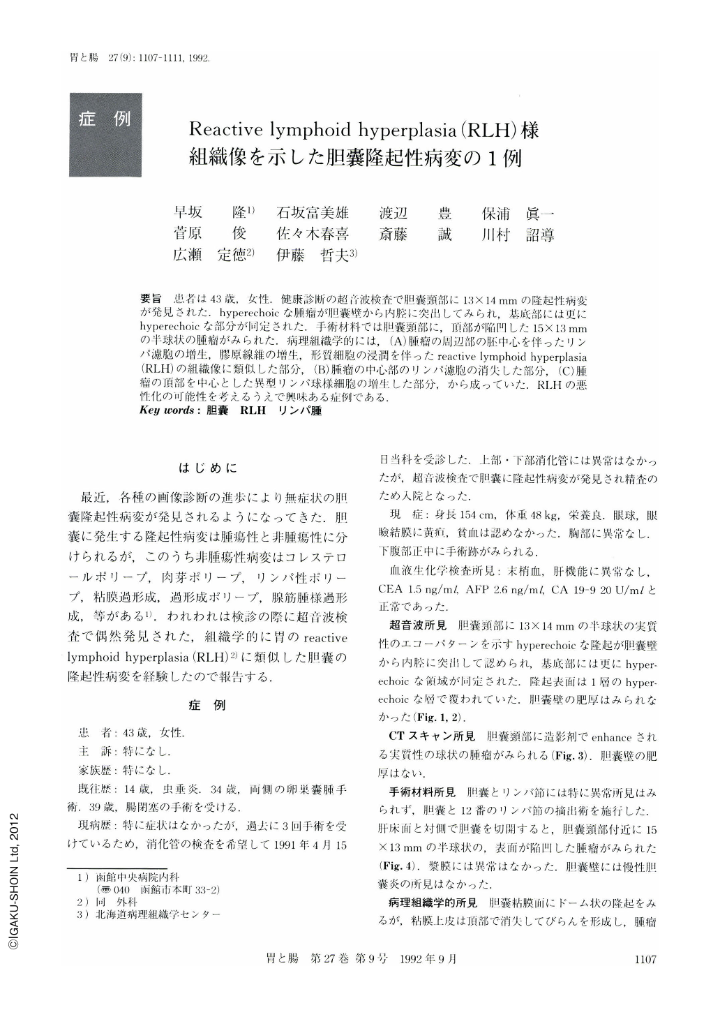
要旨 患者は43歳,女性.健康診断の超音波検査で胆囊頸部に13×14mmの隆起性病変が発見された.hyperechoicな腫瘤が胆囊壁から内腔に突出してみられ,基底部には更にhyperechoicな部分が同定された.手術材料では胆囊頸部に,頂部が陥凹した15×13mmの半球状の腫瘤がみられた.病理組織学的には,(A)腫瘤の周辺部の胚中心を伴ったリンパ濾胞の増生,膠原線維の増生,形質細胞の浸潤を伴ったreactive lymphoid hyperplasia(RLH)の組織像に類似した部分,(B)腫瘤の中心部のリンパ濾胞の消失した部分,(C)腫瘤の頂部を中心とした異型リンパ球様細胞の増生した部分,から成っていた.RLHの悪性化の可能性を考えるうえで興味ある症例である.
A 43-year-old woman was admitted to Hakodate Chuo Hospital because of a polypoid lesion of the gallbladder. A hyperechoic mass in the gallbladder was identified by ultrasonography. The resected specimen showed a hemispherical tumor with central depression. Histopathologically, the tumor consisted of three areas: 1) RLH-like lesion in the surrounding area, which was composed of proliferation of lymphatic nodules, abundant collagen fibers and plasma cells. 2) Atypical lymphocytic cell proliferation in the central are aaccompanied by plasma cells with disappearance of lymphatic nodules. 3) Diffuse atypical lymphocytic proliferation in the upper area. These three areas may be contiguous. It is suggested that this RLH-like lesion might be premalignant. This case was thought to be worth reporting because of the relationship between RLH and malignant lymphoma.

Copyright © 1992, Igaku-Shoin Ltd. All rights reserved.


