Japanese
English
- 有料閲覧
- Abstract 文献概要
- 1ページ目 Look Inside
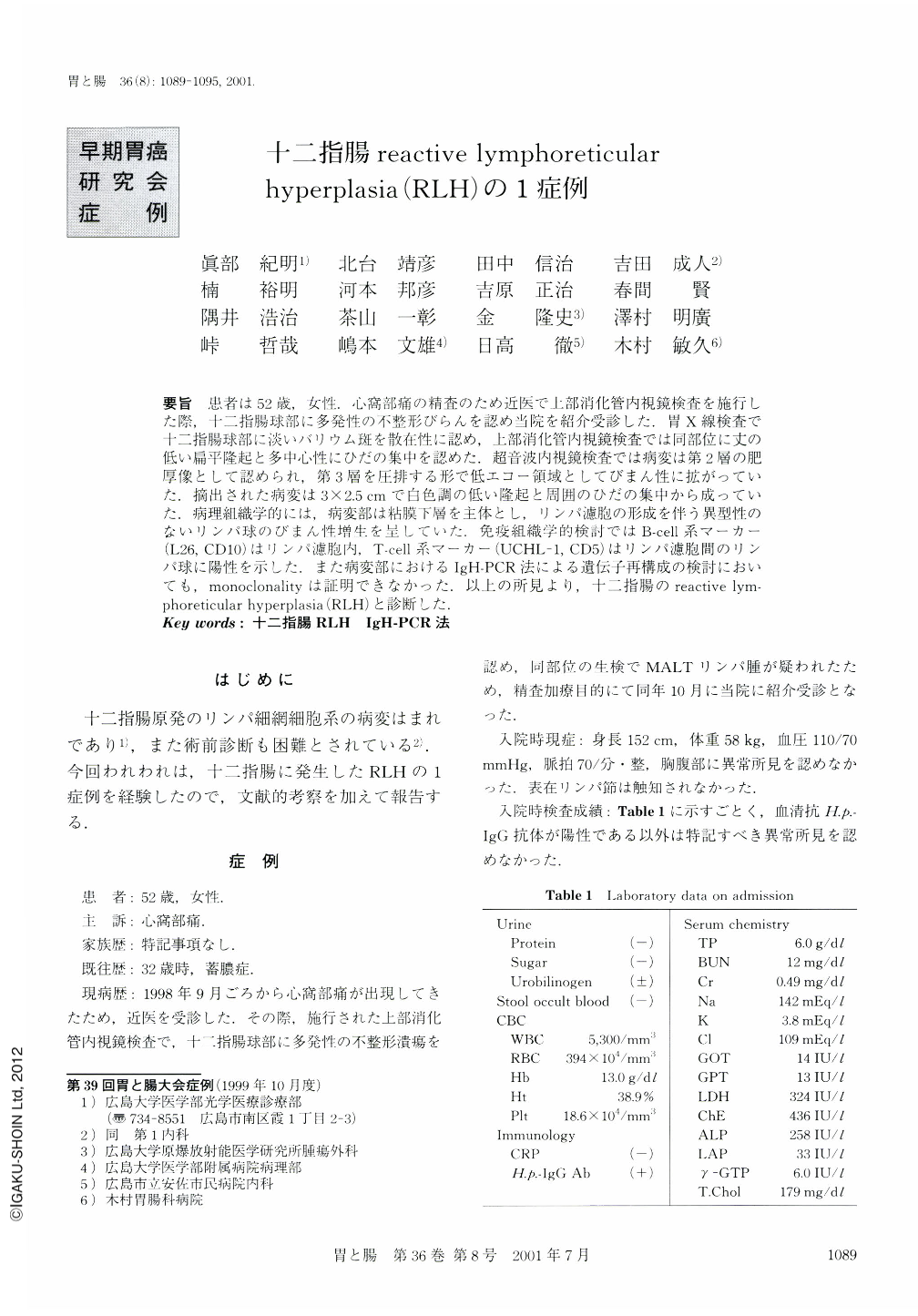
要旨 患者は52歳,女性.心窩部痛の精査のため近医で上部消化管内視鏡検査を施行した際,十二指腸球部に多発性の不整形びらんを認め当院を紹介受診した.胃X線検査で十二指腸球部に淡いバリウム斑を散在性に認め,上部消化管内視鏡検査では同部位に丈の低い扁平隆起と多中心性にひだの集中を認めた.超音波内視鏡検査では病変は第2層の肥厚像として認められ,第3層を圧排する形で低エコー領域としてびまん性に拡がっていた.摘出された病変は3×2.5cmで白色調の低い隆起と周囲のひだの集中から成っていた.病理組織学的には,病変部は粘膜下層を主体とし,リンパ濾胞の形成を伴う異型性のないリンパ球のびまん性増生を呈していた,免疫組織学的検討ではB-cell系マーカー(L26,CD10)はリンパ濾胞内,T-cell系マーカー(UCHL-1,CD5)はリンパ濾胞間のリンパ球に陽性を示した.また病変部におけるIgH-PCR法による遺伝子再構成の検討においても,monoclonalityは証明できなかった.以上の所見より,十二指腸のreactive lymphoreticular hyperplasia(RLH)と診断した.
A 52-year-old woman was admitted to our hospital for a close examination and treatment of an ulcerative lesion in the duodenal bulb. X-ray examination showed multiple small barium flecks and flat protrusions in the duodenal bulb. Endoscopic examination showed erosions, erythemas, nodular elevations, and fold convergence in the duodenal bulb. Ultrasonography showed thickening of the mucosal layer with a low echogenic level. The histology of this case was reviewed carefully and repeatedly on the suspicion of malignant lymphoma. However, the only location in which massive infiltration of the small lymphocytes with swelled lymph follicles was seen was in the duodenal bulb, and no evidence of malignancy was detected. Immunohistochemistry and molecular study concerning IgH rearrangement showed no monoclonality of the infiltrating lymphocytes. This case was diagnosed as reactive lymphoreticular hyperplasia (RLH) of the duodenal bulb.

Copyright © 2001, Igaku-Shoin Ltd. All rights reserved.


