Japanese
English
- 有料閲覧
- Abstract 文献概要
- 1ページ目 Look Inside
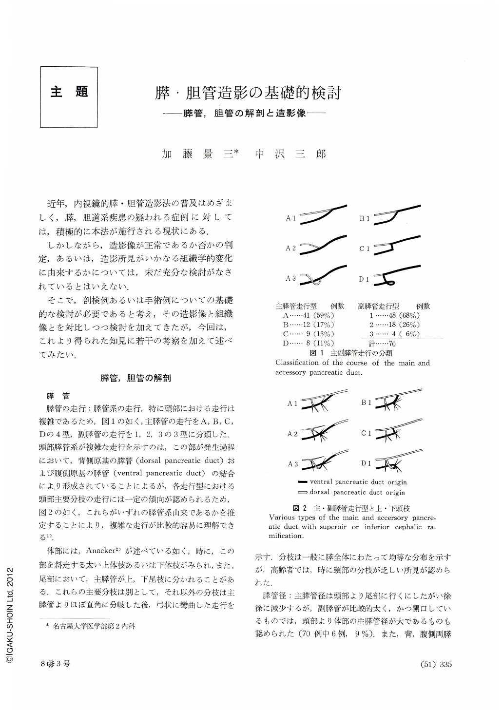
近年,内視鏡的膵・胆管造影法の普及はめざましく,膵,胆道系疾患の疑われる症例に対しては,積極的に本法が施行される現状にある.
しかしながら,造影像が正常であるか否かの判定,あるいは,造影所見がいかなる組織学的変化に由来するかについては,未だ充分な検討がなされているとはいえない.
Normal and abnormal pictures of visualized ducts of the pancreas and biliary tract have been studied on cases autopsied and operated on, and the following results have been obtained:―
A. Pancreatic ducts.
1. The courses of the major duct were classified into 4 types and those of the accessory ducts into 3. Embryologic study of those types makes it relatively easy to understand complicated courses of the duct system of the pancreas.
2. Findings such as increased diameter of the duct, its undulating contour and round, cystic dilatation of the branching ducts are closely related with changes in the epithelium of the pancreatic ducts and with intraluminal accumulation of mucus-like substance, but there is no direct relationship between these findings and those of tissue in the pancreatic parenchyma. On the other hand, rosary-shaped dilatation of the major duct and irregular, cystic dilatation of the branching ducts are both considered to reflect histologic changes in the parenchyma of the pancreas.
3. Cancer of the pancreas shows stricture and obstruction of the major duct accompanied with irregular outlines as well as cessation or straightening of the branching ducts. These findings are directly related with the question to what degree the wall of the pancreatic ducts is infiltrated with cancer. And it has been shown that striking proliferation of the connective tissue that accompanies cancer involvement is most responsible for it. The ducts caudal from the stricture are dilated.
B. Common bile duct.
1. The relationship between the bile duct and pancretic duct greatly varies according to individuals, but in the majority of cases, some connection is seen.
2. Irregular outline of the duct is caused by papillary proliferation of the epithelium of the bile duct.
3. Most cases of the dilated duct show histologic changes in the papillary region.
4. Stricture of the bile duct is not a mere reflection of histologic changes in the pancreas. A most contributing factor for it is hyperplasia of the connective tissue between the bile duct and pancreas and in the wall of the bile duct.
5. In cancers of the pancreas and bile duct uneven stricture with irregular contour is seen together with narrowing or obstruction of the duct. Not only cancerous proliferation but also hyperplasia of the tissue around the bile duct along with papillary proliferation of the epithelium of the bile duct are involved in the stricture or obstruction.
Copyright © 1973, Igaku-Shoin Ltd. All rights reserved.