Japanese
English
- 有料閲覧
- Abstract 文献概要
- 1ページ目 Look Inside
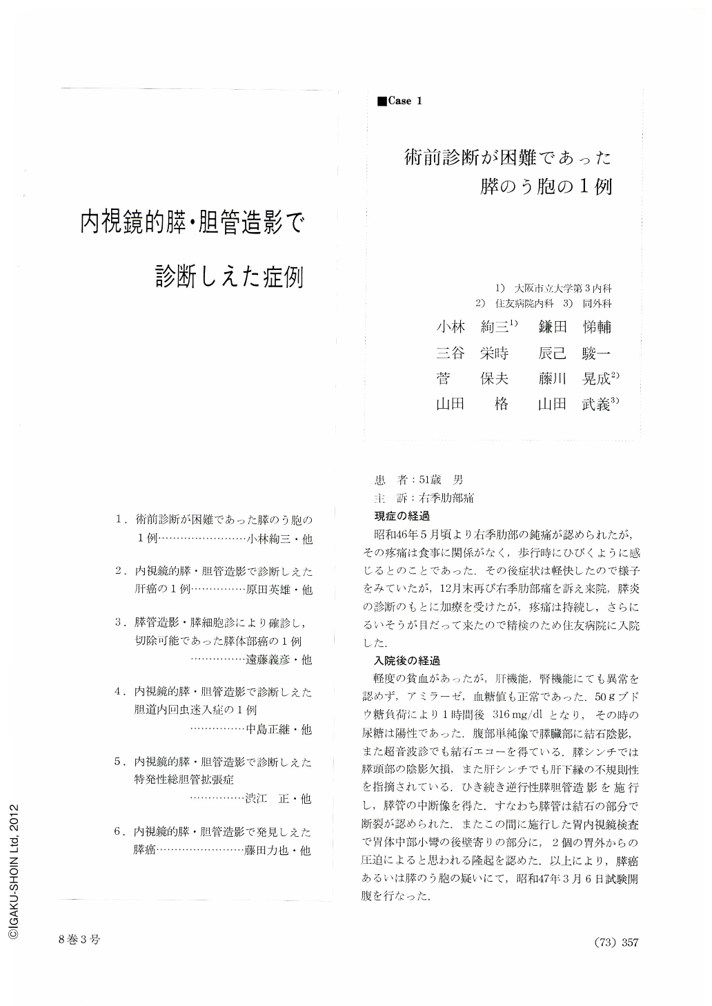
患者:51歳男
主訴:右季肋部痛
現症の経過
昭和46年5月頃より右季肋部の鈍痛が認められたが,そのとう痛は食事に関係がなく,歩行時にひびくように感じるとのことであった.その後症状は軽快したので様子をみていたが,12月末再び右季肋部痛を訴え来院,膵炎の診断のもとに加療を受けたが,とう痛は持続し,さらにるいそうが目だって来たので精検のため住友病院に入院した.
Case: a 51-year-old man.
Chief complaint: pain in the right upper quadrant of the abdomen.
Course of the present illness: Since May 1971 he felt pain in the right upper quadrant. It was unrelated to eating, but he felt it whenever he walked.
However, he did not seek any medical help because the pain had subslded for a while. At the end of December he revisited our hospital complaining of pain in the right upper quadrant. He was treated under a diagnosis of pancreatitis, but the pain persisted and weight loss was so considerable that he was admitted to Sumitomo Hospital for a thorough check-up.
Course in the hospital: Slight anemia was noticed, but otherwise no abnormality was seen in liver and kidney function tests, nor was there any abnormal levels of amylase and glucose in the blood. GTT with 50 grams of sugar showed 316 mg per 100 ml after one hour. The urine was positive for sugar then. Plain x-ray film of the abdomen revealed a shadow of a stone in the area of the pancreas. By ultrasonic waves a stone echo was also obtained.
Scintiscanning of the pancreas showed a shadow defect in the head, while in that of the liver irregularity of the lower margin was noticed. Subsequent retrograde pancreatocholangiography revealed the pancreatic duct interrupted at the site of the stone, Endoscopy of the stomach done in the interim disclosed two protrusions, probably due to extragastric compression, located on the lesser curvature of the midbody slightly on the side of the posterior wall.
On March 6, 1972, the patient underwent exploratory laparotomy under a provisional diagnosis of either of cyst of the pancreas.
Discussion:
We did not notice any icterus, and biochemical study of the blood hardly contributed to adiagnosis of pancreatic cancer. Nevertheless, we could not completely rule out cancer, because he had persistent pain and considerable loss of weight. Besides, ductal interruption was noticed in x-ray films. Cancer was finally ruled out by the operation, and multiple pancreatic cysts were demonstrated. Any connection between the cysts and the major duct was not clarified, but even if there had been any, the cysts could not have been visualized because of stone. Retrograde pancreatography is great merit in the diagnosis of pancreatic cancer, but nevertheless many false positive cases are possibly at hand. In this respect, more minute reading of opacified pancreatic duct is required hereafter.

Copyright © 1973, Igaku-Shoin Ltd. All rights reserved.


