Japanese
English
- 有料閲覧
- Abstract 文献概要
- 1ページ目 Look Inside
腸結核は,近年抗結核剤の発達に伴い,その頻度は減少してきているが,本邦では決してまれな疾患でなく,しばしば経験されている.なかでも,孤在性腸結核の占める比率は最近増大する傾向にある.特に本邦においては,散見されるクローン病との鑑別が,大きな問題となってきている.
今回,われわれは明らかな潰瘍形成や潰瘍瘢痕を認めない,興味ある肉眼病理所見を呈した回盲部腸結核の1例を経験したので報告する.
We experienced a case of ileocecal tuberculosis with a distinctive macroscopic feature.
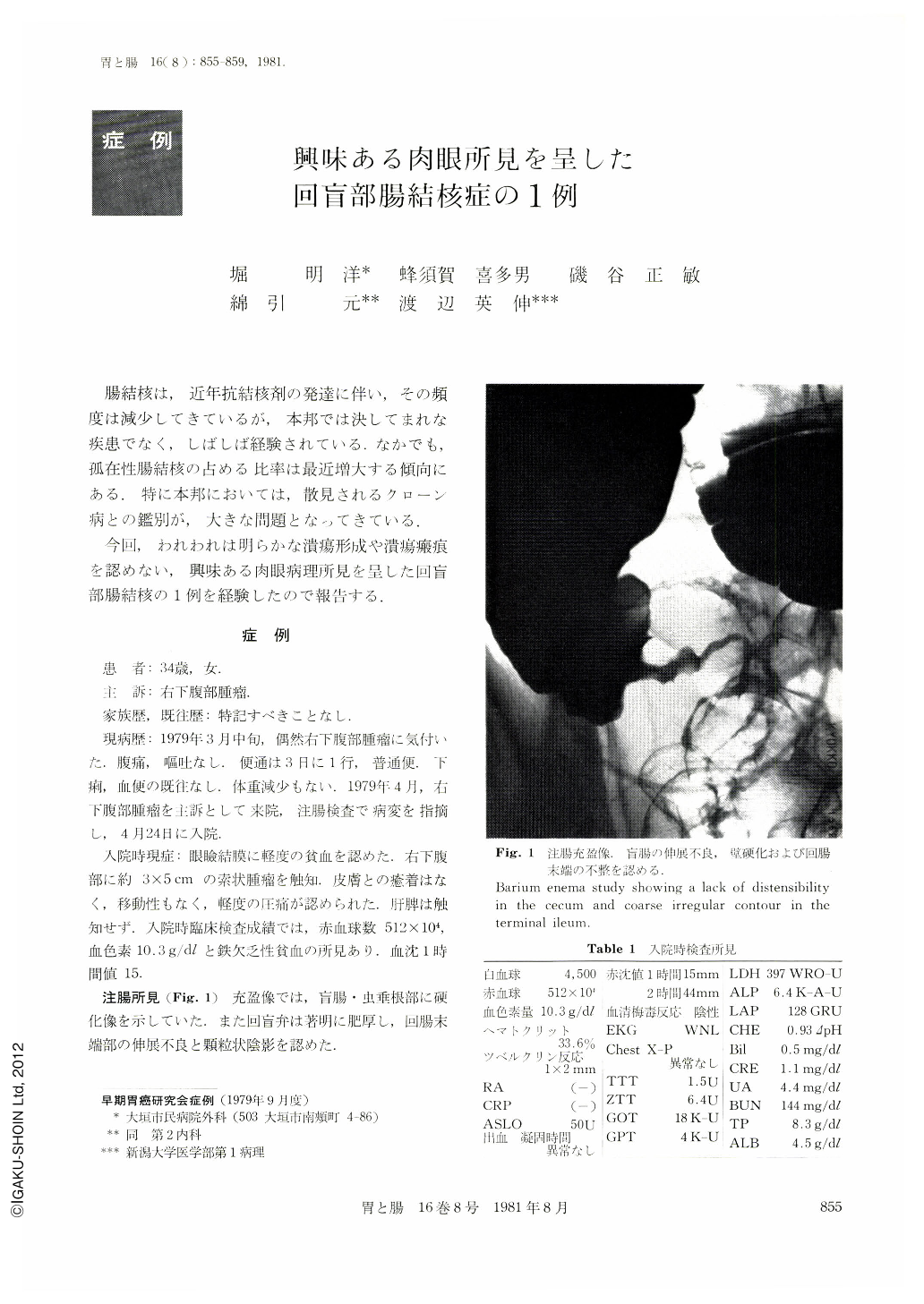
A woman aged 34 complained of a right lower abdominal mass. X-ray examination of the chest revealed no abnormalities. Barium enema study revealed a lack of distensibility in the cecum and coarse irregular contour in the terminal ileum. Double contrast study of the terminal ileum showed a coarse granular pattern of the mucosa. Endoscopic study of the colon and the terminal ileum by dye scattering method (Evans' blue) clarified multiple granular lesions. Biopsy was performed and we could find a granuloma with giant cells of Langhans'type. Terminal ileum and cecum were resected with anastomosis of the ileum and ascending colon.
Coarse granular lesions of various size were found in the terminal ileum of the resected specimen. However we could not find any ulcer or scar. In histological study the mucosa and the submucosa were swelled because of granulomas. There were caseating necrosis in some granulomas and we diagnosed this lesion as ileocecal tuberculosis. We could find no ulcer in microscopic study.

Copyright © 1981, Igaku-Shoin Ltd. All rights reserved.


