Japanese
English
- 有料閲覧
- Abstract 文献概要
- 1ページ目 Look Inside
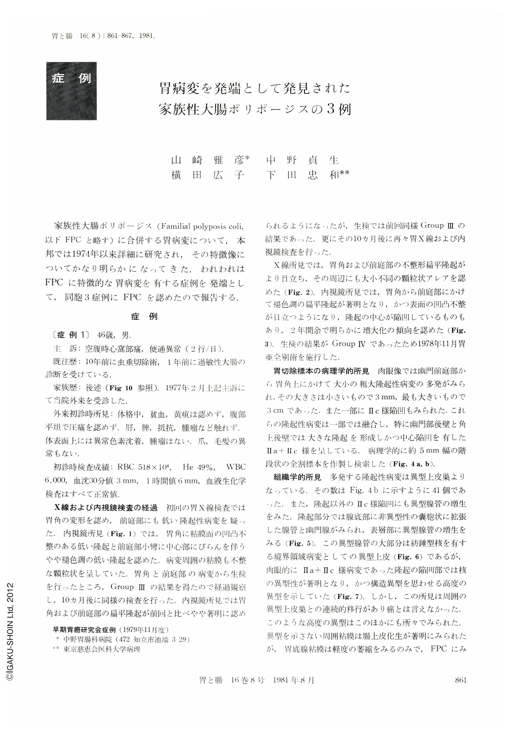
家族性大腸ポリポージス(Familial polyposis coli,以下FPCと略す)に合併する胃病変について,本邦では1974年以来詳細に研究され,その特徴像についてかなり明らかになってきた.われわれはFPCに特徴的な胃病変を有する症例を発端として,同胞3症例にFPCを認めたので報告する.
The first patient, a 46-year-old man, presented to our clinic on February 15, 1977, with complaints of hunger epigastralgia and passing of loose stool for ten days. Physical examination showed no abnormality.
Laboratory investigations were all within normal limits. X-ray and endoscopic examinations of the stomach performed in February 1977 revealed an irregular shaped flat protrusion in the gastric angle and a slightly discolored elevation with central erosion in the antrum. They were histologically diagnosed as atypical epithelium. Ten months later these gastric lesions became remarkable, but their histological findings were the same as the previous ones. Another ten months later they enlarged remarkably and mucosal surface of the antrum became rough and nodular. Histologically carcinoma was highly suspected. On November 27, 1978, a subtotal gastrectomy was performed. The gross findings of the resected specimen showed roughly nodular or irregularly circular elevated lesions of various size in an extended area from the angle to the prepploric region. Some of them fused mutually and formed large protrusions especially in the posterior wall of the antrum and above the angle. Some of the fused lesions had central depressions and formed Ⅱa+Ⅱc-like lesions. Pathologic examination revealed multiple lesions of atypical epithelium, which were located not only on the elevated regions but also on the flat ones. No precise cancerous lesion could be found. This time the patient was suspected to have familial polyposis coli. Barium enema examination and colonofiberscopy showed 27 polyps in the entire colon. Some of them were endoscopically excised.
Almost all of these resected polyps were histologically composed of tubular adenoma, but one of them had carcinoma in adenoma. X-ray examination of the small intestine and orthopantomography showed no abnormality. Duodenal mucosa was histologically normal.
The second patient, a 54-year-old man, a brother of the first patient has been followed up because of atypical epithelium on the anterior wall of the gastric angle since 1972. On July 15, 1974, he underwent abdominoperineal resection for the advanced cancer of rectum. Five years later three polyps were found in the residual colon and endoscopically resected. They were histologically diagnosed as tubular adenoma. Barium meal examination of the small bowel and orthopantomography showed normal results.
The third patient, a 49-year-old man, another brother of the first, visited our clinic on November 2, 1978, for a complete examination of the G. I tract. Physical examination showed no abnormality. Laboratory studies gave normal results. X-ray and endoscopic examinations of the stomach and colon were performed. Twenty four polyps could be found in the entire colon. Some of them were endoscopically polypectomised and histologically composed of tubular adenoma. X-ray examination of the small bowel and orthopantomography revealed no abnormality.
Family history of these three cases: Maternal grandmother died of cancer. Mother died of gastric cancer. A female sibling died at 21 of ileus due to unknown cause.

Copyright © 1981, Igaku-Shoin Ltd. All rights reserved.


