Japanese
English
- 有料閲覧
- Abstract 文献概要
- 1ページ目 Look Inside
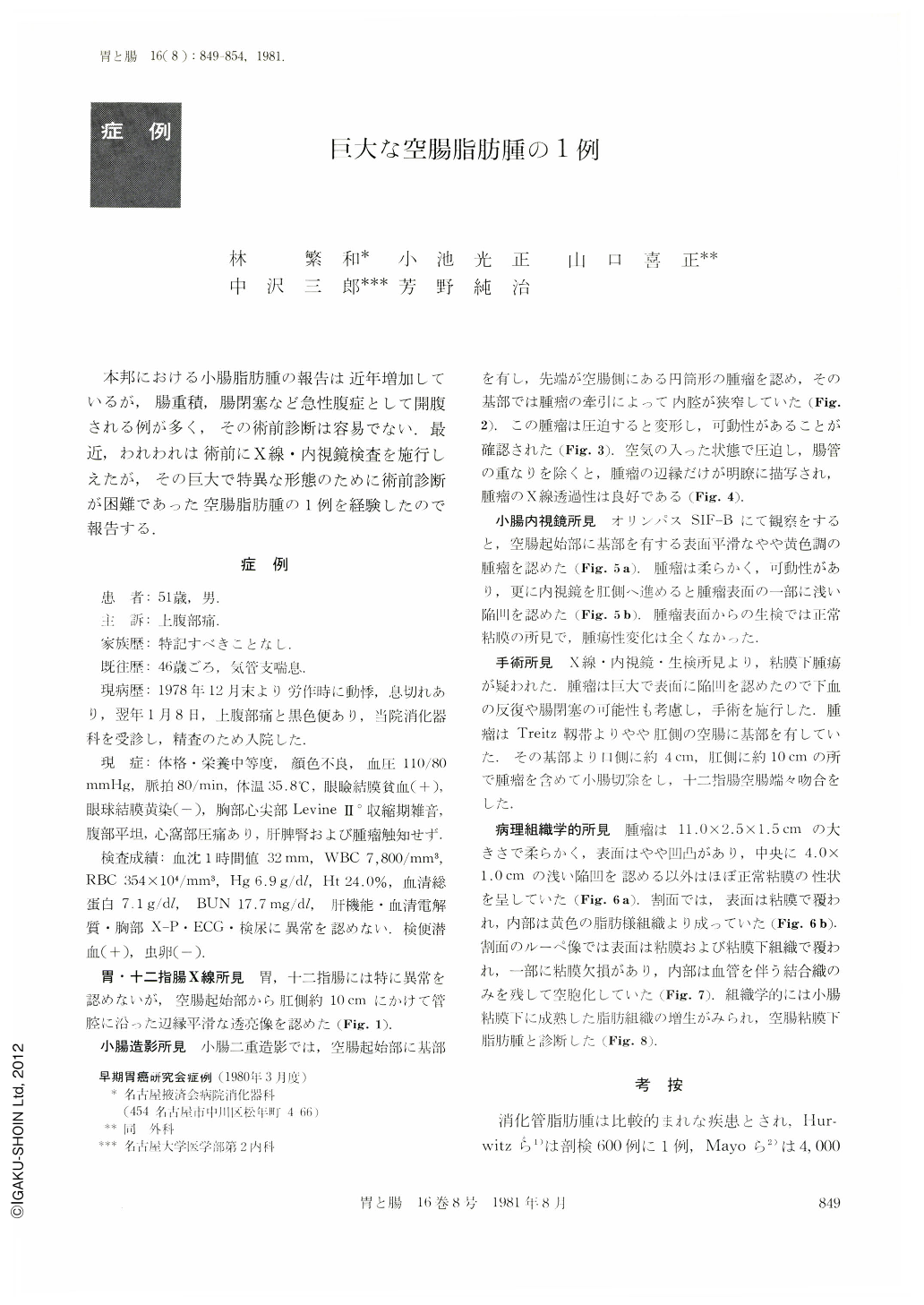
本邦における小腸脂肪腫の報告は近年増加しているが,腸重積,腸閉塞など急性腹症として開腹される例が多く,その術前診断は容易でない.最近,われわれは術前にX線・内視鏡検査を施行しえたが,その巨大で特異な形態のために術前診断が困難であった空腸脂肪腫の1例を経験したので報告する.
A 51-year-old man was admitted to our hospital with a major complaint of epigastralgia. Through ordinary examinations, none other than hypochromic anemia and positive occult blood of the stool were seen. By x-ray of the small intestine, a movable smooth cylindrical tumor with the head at the jejunum and the base in the beginning portion of the jejunum was found. Because the tumor changed its shape with compression, radiolucency was satisfactory. Endoscopy of the small intestine showed a yellowish tumor with a smooth even surface and a shallow depression in one part. Biopsy showed no malignancy.
According to these results, it was supposed as subnnicosal tumor and due to its large size and depreslion at its surface, possibility of repeated bleeding and ileus was taken into consideration and resection of the small intestine was perfornled subsequently. The base of the tumor was situated more on the anal side of the jejunum than the Threitz ligament,11.0×2.5×1.5cm in size, soft, and the mucosa in a close to normal sate other than a 4×1cm narrow deprespion in the center. The cut surface was yellow with fatty tissue. Histologically, increased growth of mature adipose tissue in the submucosal layer was seen. 108 cases of lipoma of the small intestine are reported in Japan, and in approximately 65%, they are complicated with invagination and ileus which makes preoperative diagnosis difficult. There are 41 cases of those which underwent x-ray examination but only 25 show the lesion and of which seven are of the duodenum. Our case is the only reported one of the jejunum. Lipoma shows excellent radiolucency showing changes with pressure but diagnosis with x-ray examination is not easy. There are only eleven cases of endoscopical observations and this report is the first of observations in Japan of the small intestine eticluding the duodenum. Because this case showed no complication with invagination, x-ray and endcscopy could be performed, but preoperative diagnosis was difficult due to its large size and unique charac[enstics.

Copyright © 1981, Igaku-Shoin Ltd. All rights reserved.


