Japanese
English
- 有料閲覧
- Abstract 文献概要
- 1ページ目 Look Inside
症 例
患 者:45歳 男性
主 訴:悪心,嘔吐,腹部膨満
家族歴・既往歴:特記すべきものなし
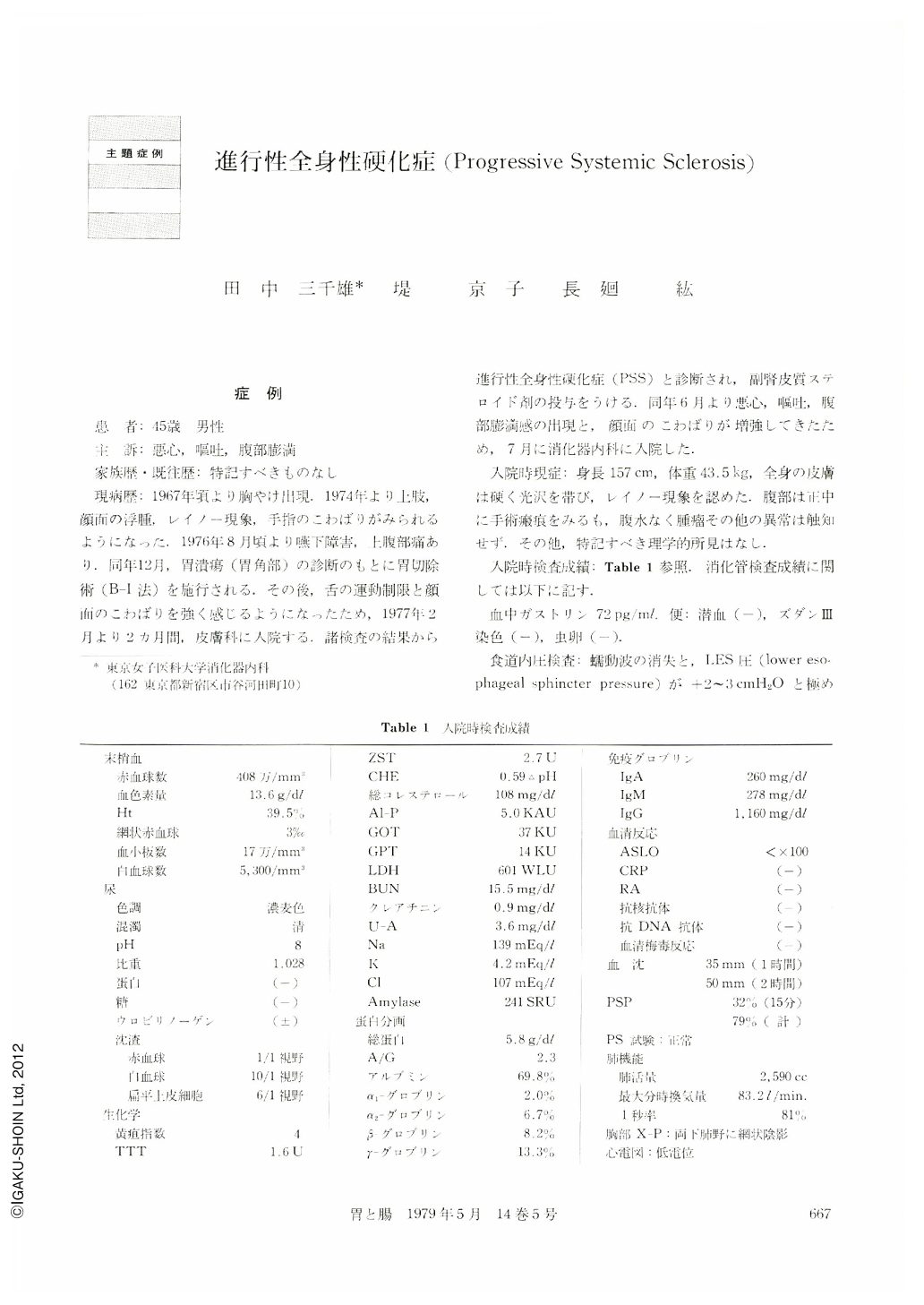
現病歴:1967年頃より胸やけ出現.1974年より上肢,顔面の浮腫,レイノー現象,手指のこわばりがみられるようになった.1976年8月頃より嚥下障害,上腹部痛あり.同年12月,胃潰瘍(胃角部)の診断のもとに胃切除術(B-Ⅰ法)を施行される.その後,舌の運動制限と顔面のこわばりを強く感じるようになったため,1977年2月より2カ月間,皮膚科に入院する,諸検査の結果から進行性全身性硬化症(PSS)と診断され,副腎皮質ステロイド剤の投与を受ける.同年6月より悪心,嘔吐,腹部膨満感の出現と,顔面のこわばりが増強してきたため,7月に消化器内科に入院した.
A 45-year-old man was admitted to the hospital on July 1977. At the age of 35, he noted heart burn, and at the age of 42, tightness and thickness of his skin and Raynaud's phenomenon began. Meanwhile, he developed dysphagia and epigastralgia. In Dec. 1976, gastric ulcer was found and subtotal gastrectomy (B-1) was performed, with relief of epigastralgia. Five months before admission, skin biopsy was done and diagnosis of PSS had been made. Present chief complaints were nausea, vomiting and abdominal distension.
Physical Examination-The patient was thin. The skin was thin, taut and atrophic. The hands appeared claw-like with loss of extension and flexion of the fingers. The remainder of the physical examination was noncontributory.
Laboratory Data-Hb 13.6 g/100 ml, WBC 5,300, with normal differential count; blood chemistry study was essentially normal, ESR 35 mm in the first hour. EKG showed low voltage. The 4-days faecal out put of 131I-Triolein was 5.9% of the parentally administered dose.
Roentgenologic Studies-Examination with barium showed dilatation of the esophagus and small intestine. Peristalsis was diminished.
Intraluminal pressures of Esophagus-Disappearance of a peristaltic wave and low pressure (+2~3 cm H2O) at the lower esophageal sphincter were demonstrated.
Endoscopic studies-Mucosal redness was observed in the lower part of esophagus and duodenum.
Biopsy on the duodenum-Atrophy of the villi and capillary dilatation were recognized.
Course-Treatment was performed symptomatically. The patient became progressively weaker and died eight month after entry.
Autopsy-The esophagus and the duodenum were slightly dilated, and the jejunum was makedly dilated. The mucosa of the lower esophagus and the duodenum was hyperemic.
Histological Examination-All sections from all parts of the alimentary tract showed diffuse atrophy of the muscularis and increased deposition of collagen in the submucosa and muscular layer. Vascular abnormality, such as fibrosis and concentric intimal proliferation, was not recognized in all parts of the gastrointestinal tract.
Comment-PSS is recognized as a member of the so-called collagen group of diseases. Involvement of the gastrointestinal tract in PSS has received increasing attention. The pathological changes consist of a systemic alteration in the connective tissue and vascular system. This patient's gastrointestinal symptoms was severe. Abdominal distension, disphagia, nausea and vomiting dominated the clinical picture. Dilatation of the esophagus and small intestine, diminution of peristalsis and malabsorption were demonstrated by the clinical examination. Histological study showed atrophy and partial replacement of the muscular coat by collagenous tissue in all part of the alimentary tract. But vascular alteration of the G-I tract was not recognized. Autonomic nervous system dysfunction was suggested to be one of the pathogenesis of Raynaud's phenomenon in PSS. The same phenomenon without morphological changes of the blood vessel may exist at the G-I tract.

Copyright © 1979, Igaku-Shoin Ltd. All rights reserved.


