Japanese
English
- 有料閲覧
- Abstract 文献概要
- 1ページ目 Look Inside
Peutz-Jeghers症候群は1921年Peutz1)によりはじめて記録され,1949年Jeghersら2)によって確立された口唇,口腔粘膜,指趾の色素斑,および胃,腸ポリポージス,単性優性遺伝を示す疾患である.最近われわれは腸重積症を併発したこの典型的な1症例を経験したので報告し,あわせて若干の文献的考察を試みる.
症例
患 者:11歳 女子 小学生
主 訴:腹痛及び嘔吐
既往歴:2歳頃より口唇に色素斑を認め,1973年1月に腹痛,嘔吐を認めた.
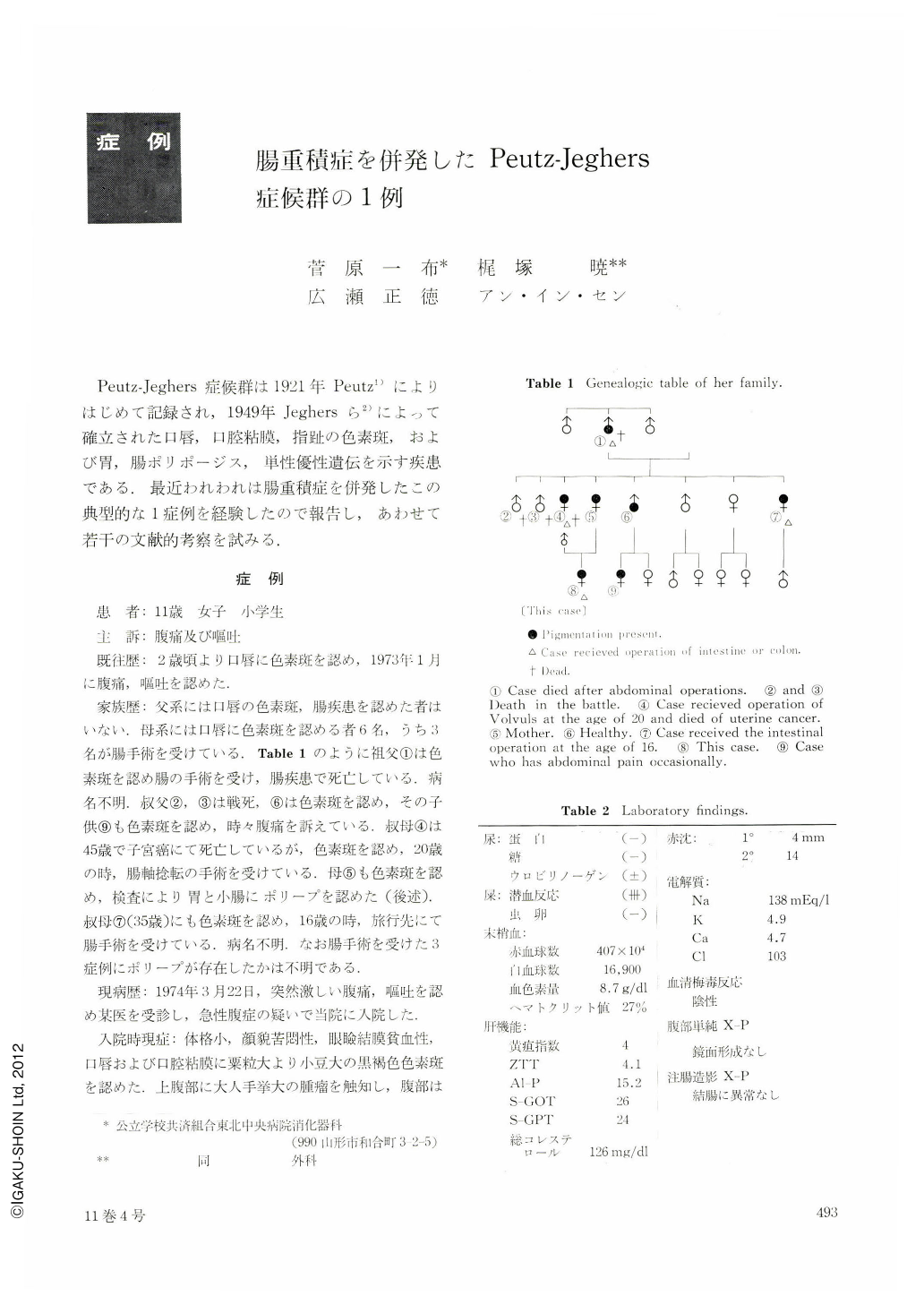
家族歴:父系には口唇の色素斑,腸疾患を認めた者はいない.母系には口唇に色素斑を認める者6名,うち3名が腸手術を受けている,Table1のように祖父①は色素斑を認め腸の手術を受け,腸疾患で死亡している,病名不明.叔父②,③は戦死,⑥は色素斑を認め,その子供⑨も色素斑を認め,時々腹痛を訴えている.叔母④は45歳で子宮癌にて死亡しているが,色素斑を認め,20歳の時,腸軸捻転の手術を受けている.母⑤も色素斑を認め,検査により胃と小腸にポリープを認めた(後述).叔母⑦(35歳)にも色素斑を認め,16歳の時,旅行先にて腸手術を受けている.病名不明.なお腸手術を受けた3症例にポリープが存在したかは不明である.
現病歴:1974年3月22日,突然激しい腹痛,嘔吐を認め某医を受診し,急性腹症の疑いで当院に入院した.
入院時現症:体格小,顔貌苦悶性,眼瞼結膜貧血性,口唇および口腔粘膜に粟粒大より小豆大の黒褐色色素斑を認めた.上腹部に大人手挙大の腫瘤を触知し,腹部は鼓音を呈した.手掌,足底および指先にも黒褐色色素斑を認めた(後述).下血は認めない.
検査成績:便の潜血反応が強陽性であった.腹部単純撮影では鏡面形成みられず,注腸造影でも結腸に異常は認めなかった(Table 2).
入院後経過:入院後も腹痛,嘔吐は続き,入院第3病日に施行した涜腸後,多量の1血便がみられた.入院第4病日に小腸重積症の診断のもとに開腹手術を行なった.
The patient is a 11-year-old girl. Family history revealed that there are six individuals with pigmentation on the lips in her maternal side and three of them underwent intestinal operation. The patient has had pigmentation spots on the lips since the age of two. On March 22, 1974, she suddenly had a severe abdominal pain accompanied by nausea and vomiting, and was admitted to the Tohoku Chuo Hospital as acute abdomen. Physical examination on admission showed that a number of dark-brown colored pigmentation spots ranging from a millet grain to red bean in size were noted on the lips, buccal mucosa, palms of the hands and soles the feet. A tumor mass about the size of fist was palpated in the upper abdomen.
Although she had no episode of melena, stools were strongly positive for occult blood. Both the plain film of the abdomen in upright position and examination of the colon by barium enema revealed no abnormal finding.
Four days after admission, a surgical intervention revealed that, the tumor mass palpated was found to be intussusception of the jejunum located about 12 cm distal to the Treit'z ligament. About 80 cm of the involved area was resected. Four polyps were detected in the removed specimen. One of them was cauliflower-shaped and about 2 cm in diameter. Since this polyp had a stalk, it was postulated that the indigitation was caused by prolapse of this pedunculated tumor. Histologically, this was found to be hyperplastic polyp. Moreover, examination of upper GI tract revealed another polyp 1. 5 cm in diameter located in the body of the stomach. It is also noteworthy that her mother has pigmentation spots on the lips, buccal membrane, palms of the hands and soles of the feet, accompanied by gastric and small intestinal polyp.

Copyright © 1976, Igaku-Shoin Ltd. All rights reserved.


