Japanese
English
- 有料閲覧
- Abstract 文献概要
- 1ページ目 Look Inside
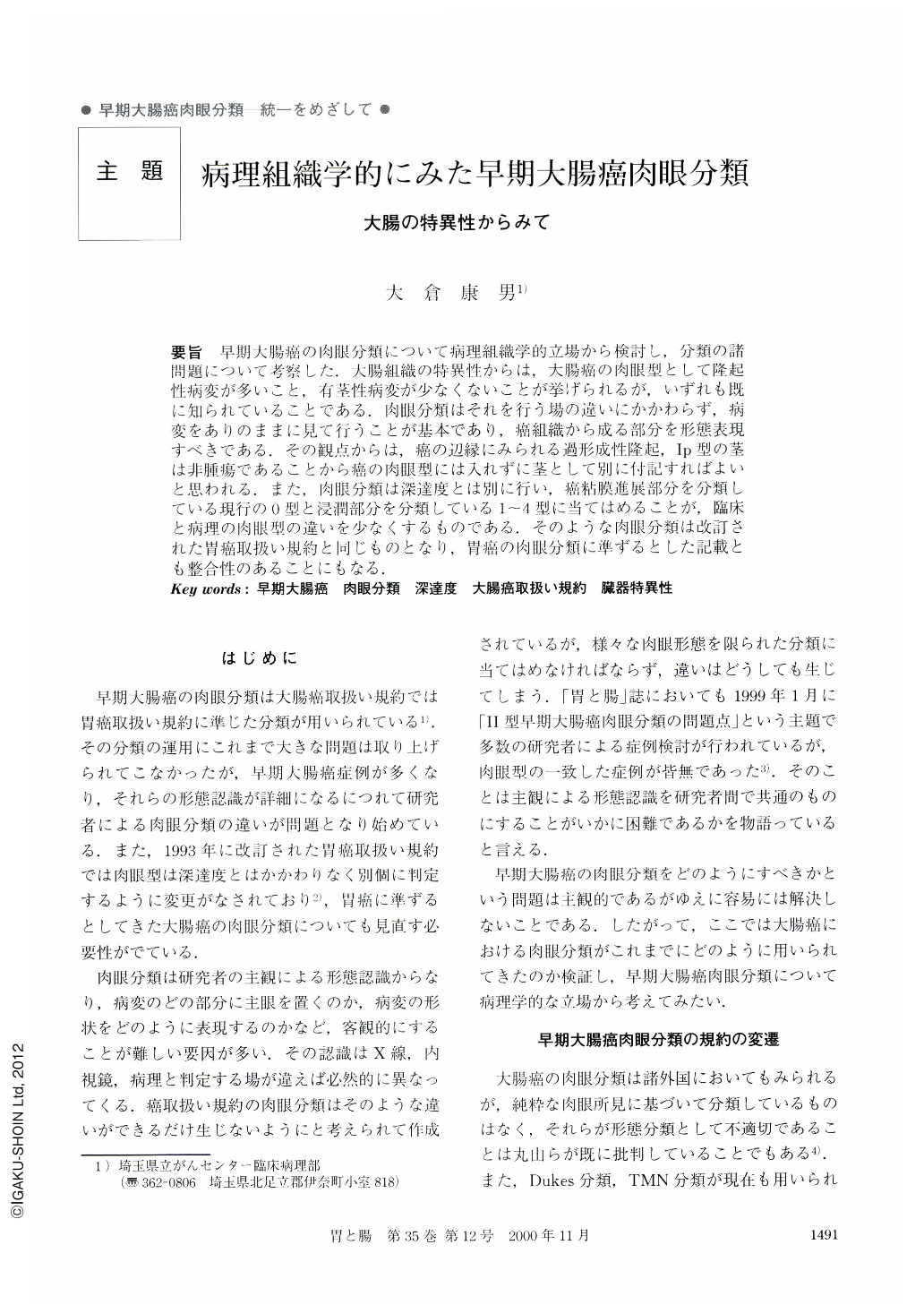
要旨 早期大腸癌の肉眼分類について病理組織学的立場から検討し,分類の諸問題について考察した.大腸組織の特異性からは,大腸癌の肉眼型として隆起性病変が多いこと,有茎性病変が少なくないことが挙げられるが,いずれも既に知られていることである.肉眼分類はそれを行う場の違いにかかわらず,病変をありのままに見て行うことが基本であり,癌組織から成る部分を形態表現すべきである.その観点からは,癌の辺縁にみられる過形成性隆起,Ⅰp型の茎は非腫瘍であることから癌の肉眼型には入れずに茎として別に付記すればよいと思われる.また,肉眼分類は深達度とは別に行い,癌粘膜進展部分を分類している現行の0型と浸潤部分を分類している1~4型に当てはめることが,臨床と病理の肉眼型の違いを少なくするものである.そのような肉眼分類は改訂された胃癌取扱い規約と同じものとなり,胃癌の肉眼分類に準ずるとした記載とも整合性のあることにもなる.
Along with the increase in number of detected early colorectal carcinomas, several problems in typing the macroscopic features have occurred. Examples are the difference between 0-Ⅱa type and 0-Ⅰs type, the naming of 0-Ⅰs type with cancerous depression, and the change of macroscopic type according to invasive depth between the submucosa and the proper muscle, and so on. To resolve these problems, the macroscopic classification of early colorectal carcinomas was reexamined from the histopathological view point of organ specificity.
The layer of mucosa and muscularis mucosa in the colonic wall is thinner than that in the gastric wall. Because of this specific difference between these two organs, carcinoma in the colorectum tend to have longer stalks. Moreover, judging from the frequency of tumors of glandular structure, it can be seen that protruded lesions in the colorectum are more numerous than in the gastric wall. Clinicopathologically, these features are already recognized.
To unify the macroscopic type found in barium, endoscopic and pathological examinations, the classification was made by the cancerous figures done. Because of this, the protruded mucosa of reactive hyperplasia which is usually found around the carcinoma was not included among the features of the macroscopic type. For the same reason, the stalk of the 0-Ⅰp type, most of which is constructed by elongated normal mucosa, was described as an additional feature along with the type of cancerous lesion.
No distinction of macroscopic type has been recognized between carcinoma deeply invading the submucosa and carcinoma slightly invading the proper muscle. Abiding by the general rules (provided by the Japanese Society for Cancer of the Colon and Rectum) for clinical and pathological studies concerning cancer of the colon, rectum and anus, if a carcinoma which was designated as 2 type remained in the submucosa, the macroscopic type was given as 0-Ⅱa + Ⅱc type. The same problem arose when trying to distinguish between 0-Ⅰs type submucosal invasive carcinoma and 1 type advanced carcinoma. In other words, the macroscopic classification of carcinoma was decided only by the macroscopic figures without reference to the depth of invasion.
The same macroscopic classification of cancers of the colon as the new macroscopic classification of gastric carcinoma was used, so that there would be unity in classification used for the whole gastrointestinal tract.

Copyright © 2000, Igaku-Shoin Ltd. All rights reserved.


