Japanese
English
- 有料閲覧
- Abstract 文献概要
- 1ページ目 Look Inside
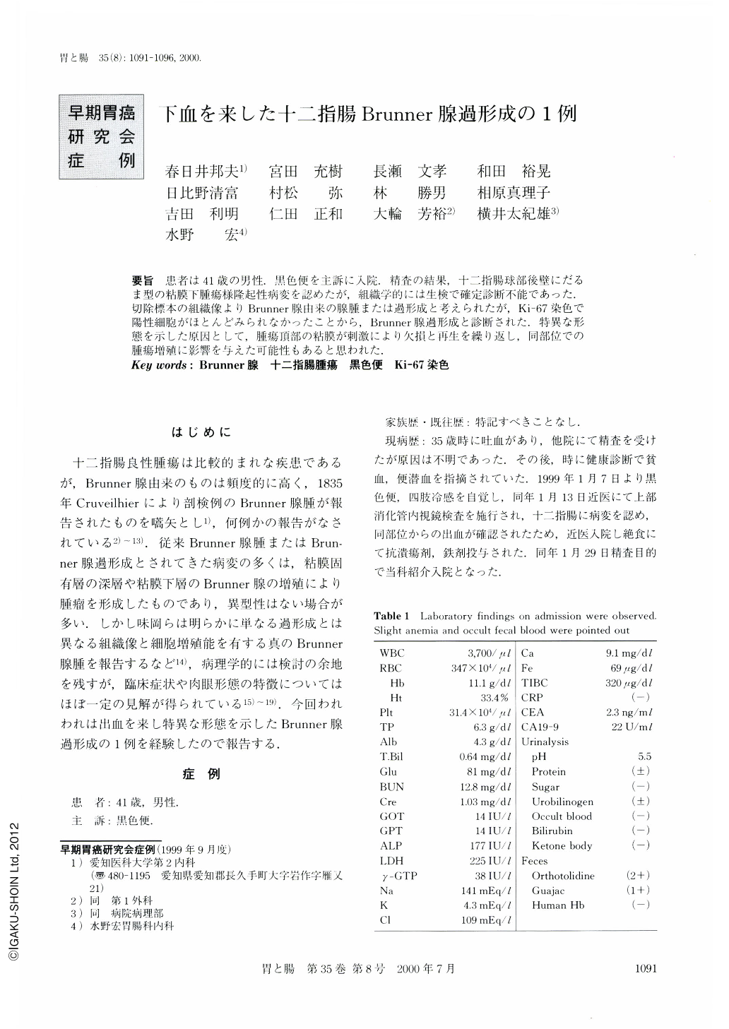
要旨 患者は41歳の男性.黒色便を主訴に入院精査の結果,十二指腸球部後壁にだるま型の粘膜下腫瘍様隆起性病変を認めたが,組織学的には生検で確定診断不能であった.切除標本の組織像よりBrunner腺由来の腺腫または過形成と考えられたが,Ki-67染色で陽性細胞がほとんどみられなかったことから,Brunner腺過形成と診断された.特異な形態を示した原因として,腫瘍頂部の粘膜が刺激により欠損と再生を繰り返し,同部位での腫瘍増殖に影響を与えた可能性もあると思われた.
A 41-year-old Japanese man was introduced to our hospital because of tarry stool. Further examination revealed a submucosal tumor-like elevated lesion in the anterior wall of the duodenal bulb. Macroscopically, the lesion appeared to have a“small half-sphere on the top” but the biopsy which was carried out in the endoscopical examination could not identify the histological cell type. The resected specimen and its histological investigation indicated that the lesion was derived from Brunner's gland, and some cells were judged as positive by immunologocal Ki-67 staining, so the lesion was finally diagnosed as Brunner's gland hyperplasia. The hypothesis was thought to possibly indicate that the curious macroscopic form of this lesion was caused by the hyperproliferation of the cells at the top of the elevated lesion. Thus may have been due to stimulation by intestinal movement and/or gastric juice.

Copyright © 2000, Igaku-Shoin Ltd. All rights reserved.


