Japanese
English
- 有料閲覧
- Abstract 文献概要
- 1ページ目 Look Inside
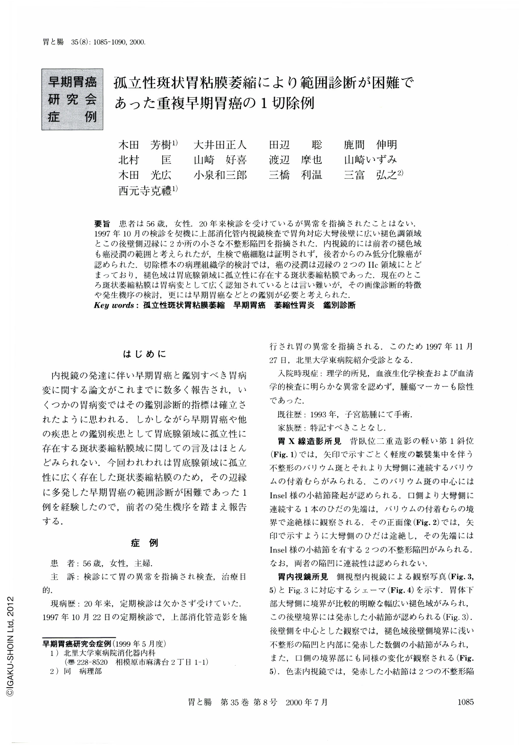
要旨 患者は56歳,女性.20年来検診を受けているが異常を指摘されたことはない.1997年10月の検診を契機に上部消化管内視鏡検査で胃角対応大彎後壁に広い褪色調領域とこの後壁側辺縁に2か所の小さな不整形陥凹を指摘された.内視鏡的には前者の褪色域も癌浸潤の範囲と考えられたが,生検で癌細胞は証明されず,後者からのみ低分化腺癌が認められた.切除標本の病理組織学的検討では,癌の浸潤は辺縁の2つのⅡc領域にとどまっており,褪色域は胃底腺領域に孤立性に存在する斑状萎縮粘膜であった.現在のところ斑状萎縮粘膜は胃病変として広く認知されているとは言い難いが,その画像診断的特徴や発生機序の検討,更には早期胃癌などとの鑑別が必要と考えられた.
An abnormality in the stomach anglus of a 56-year-old woman was pointed out during a routine medical check up. Subsequent endoscopic examination revealed a discolored area located on the posterior wall of the anglus, and double Ⅱc type lesions situated on its posterior periphery. Endoscopically, it seemed that the discolored area indicated the extent of cancer invasion of the double Ⅱc lesions. However,‘histologically' poorly differentiated adenocarcinoma cells were observed only in the biopsy specimens from the double Ⅱc lesions, and not from any points of the discolored area. Pathological examination of the resected specimen showed the discolored area was the focal atrophy of the gastric mucosa located solitarily in the fundic gland area. Both of the Ⅱc type early gastric cancers were limited to the mucosal layer, and no cancer cells extending to the discolored area were observed. In this case, the solitary focal atrophy of the gastric mucosa in the fundic gland area caused misdiagnosis of the extent of the double early gastric cancers situated on its periphery. To date, few reports have referred to the solitary focal atrophy of the gastric mucosa as an indicator for differential diagnosis of gastric cancers. The generation and endoscopic features of this lesion should be studied for differential diagnosis of early gastric cancers and other types of cancers.

Copyright © 2000, Igaku-Shoin Ltd. All rights reserved.


