Japanese
English
- 有料閲覧
- Abstract 文献概要
- 1ページ目 Look Inside
- サイト内被引用 Cited by
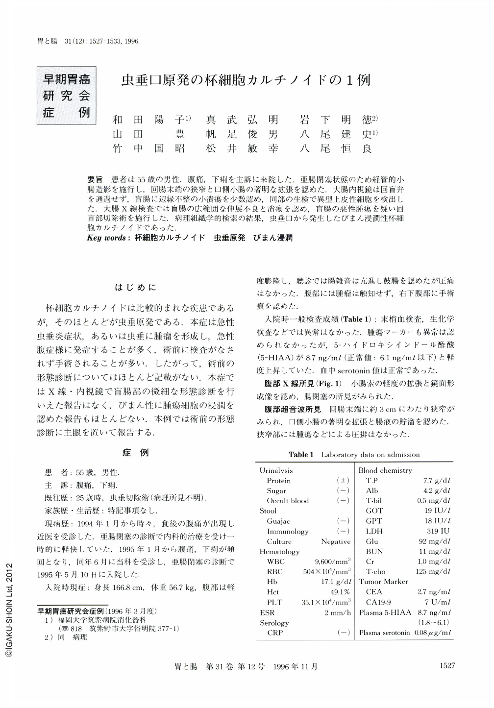
要旨 患者は55歳の男性.腹痛,下痢を主訴に来院した.亜腸閉塞状態のため経管的小腸造影を施行し,回腸末端の狭窄と口側小腸の著明な拡張を認めた.大腸内視鏡は回盲弁を通過せず,盲腸に辺縁不整の小潰瘍を少数認め,同部の生検で異型上皮性細胞を検出した.大腸X線検査では盲腸の広範囲な伸展不良と潰瘍を認め,盲腸の悪性腫瘍を疑い回盲部切除術を施行した.病理組織学的検索の結果,虫垂口から発生したびまん浸潤性杯細胞カルチノイドであった.
A 65-year-old man was admitted to our hospital with abdominal pain as his chief complaint. He had been appendectomized 30 years previously. Abdominal X-ray film showed extensive niveau formation. Retrograde selective colonography using a long rectal tube showed subtle but definite decreased extensibility of the cecum, and a small, irregular-shaped ulcer. Barium did not flow back into the ileum. Under colonoscopic examination, Bauhin's valve seemed hard and swollen and small erosions were observed around the appendiceal orifice. Histological examination of a biopsy taken from the erosions revealed atypical cells suggesting well differentiated adenocarcinoma. Because of these findings, we performed the ileocecal resection after a diagnosis of cancer of the ileocecal region. From histological examination of the resected specimen, the tumor was diagnosed as a goblet cell carcinoid which had developed from the mucosa of the appendiceal orifice and infiltrated into the cecal area mainly through the mascularis mucosa and partly through the serosa.

Copyright © 1996, Igaku-Shoin Ltd. All rights reserved.


