Japanese
English
- 有料閲覧
- Abstract 文献概要
- 1ページ目 Look Inside
- サイト内被引用 Cited by
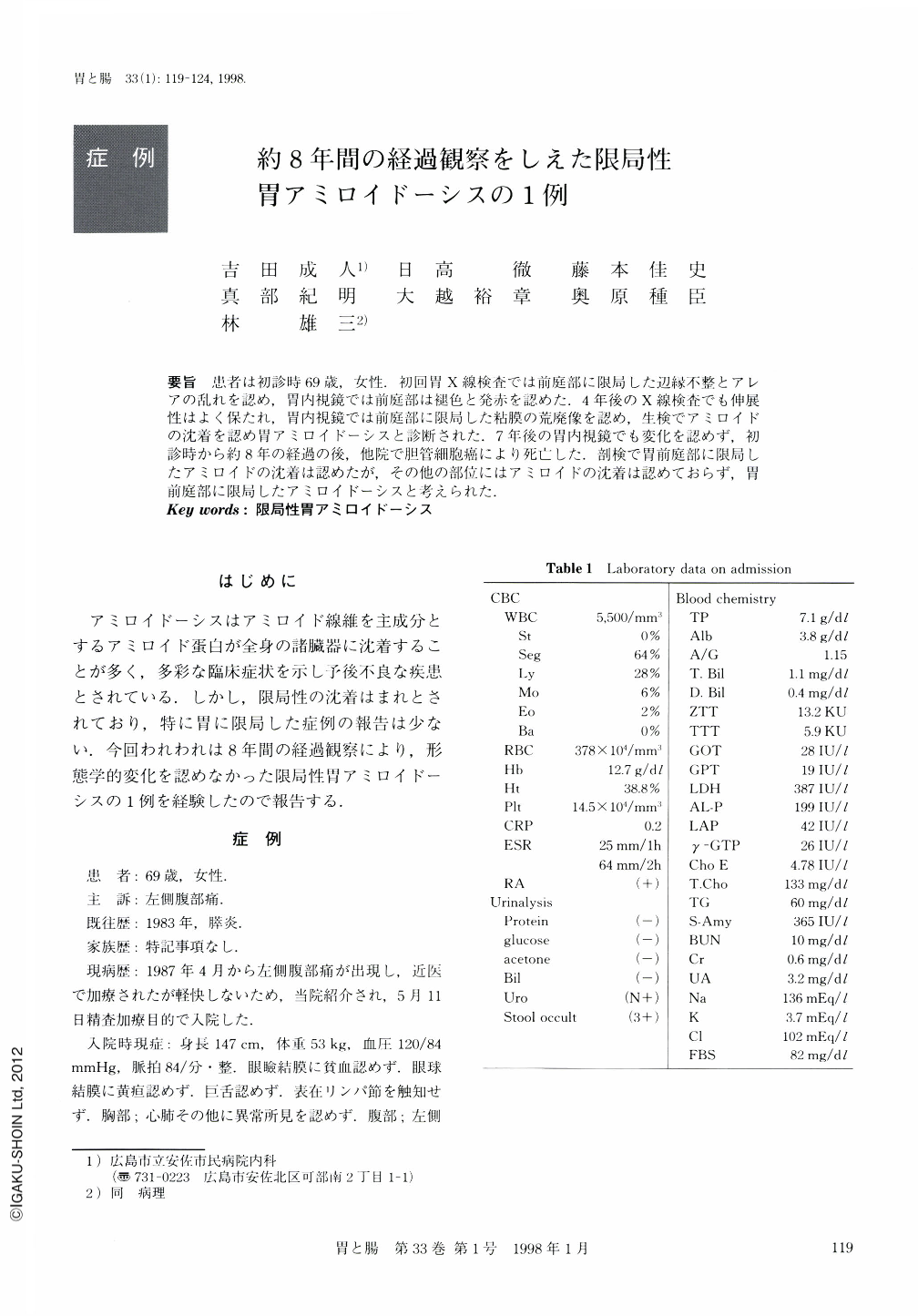
要旨 患者は初診時69歳,女性.初回胃X線検査では前庭部に限局した辺縁不整とアレアの乱れを認め,胃内視鏡では前庭部は褪色と発赤を認めた.4年後のX線検査でも伸展性はよく保たれ,胃内視鏡では前庭部に限局した粘膜の荒廃像を認め,生検でアミロイドの沈着を認め胃アミロイドーシスと診断された.7年後の胃内視鏡でも変化を認めず,初診時から約8年の経過の後,他院で胆管細胞癌により死亡した.剖検で胃前庭部に限局したアミロイドの沈着は認めたが,その他の部位にはアミロイドの沈着は認めておらず,胃前庭部に限局したアミロイドーシスと考えられた.
A 69-year-old woman was admitted to our hospital suffering from left flank pain. Radiological examination of the gastrointestinal tract showed an irregular, mucosal pattern in the antrum. Endoscopic examination revealed discoloration, redness and coarse mucosa in the antrum. Histological examination of biopsy specimens revealed no malignancy. Four years later, distension of the antrum was revealed in a radiology examination. Endoscopic examination showed a coarse mucosal pattern in the antrum, and biopsy specimens revealed amyloidosis of the stomach. Seven years later, endoscopic findings were unchanged. Eight years after the first medical examination, the patient died of cholangiocellular carcinoma at another hospital. Autopsy was performed, and histological examination showed amyloid deposits in the antrum, but not in any other organs. Based on these findings, this case was diagnosed as localized amyloidosis of the stomach.

Copyright © 1998, Igaku-Shoin Ltd. All rights reserved.


