Japanese
English
- 有料閲覧
- Abstract 文献概要
- 1ページ目 Look Inside
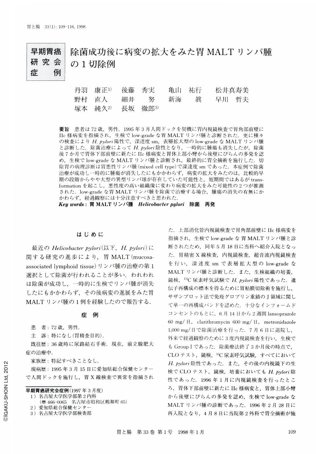
要旨 患者は72歳,男性.1995年3月人間ドックを契機に胃内視鏡検査で胃角部前壁にⅡc様病変を指摘され,生検でlow-gradeな胃MALTリンパ腫と診断された.更に種々の検査によりH. pylori陽性で,深達度sm,表層拡大型のlow-gradeなMALTリンパ腫と診断した.除菌治療によってH. pylori陰性となり,一時的に腫瘍も消失したが,除菌後7か月で胃体下部前壁に新たにⅡc様病変と胃体上部小彎から後壁にびらんの多発を認め,生検でlow-gradeなMALTリンパ腫と診断され,最終的に胃全摘術を施行した.切除胃の病理診断は胃悪性リンパ腫(mixed cell type)で深達度smであった.本症例で除菌治療が成功し一時的に腫瘍が消失したにもかかわらず,病変の拡大をみたのは,比較的早期の段階からやや大型の異型リンパ球が存在していた可能性と,短期間ではあるがtransformationを起こし,悪性度の高い組織像に変わり病変の拡大をみた可能性の2つが推測された.low-gradeな胃MALTリンパ腫を除菌で治療する場合,腫瘍の消失の有無にかかわらず,経過観察には十分注意すべきと思われた.
During a medical check-up examination, a Ⅱc-like depressed lesion was discovered in a 72-year-old man. He was admitted to our hospital for more precise examination. Radiographic and endoscopic study revealed a shallow depressed lesion with granular mucosal pattern on the anterior wall of the anglus. The histological diagnosis of the biopsy specimen revealed low grade of mucosa-associated lymphoid tissue lymphoma (MALToma). Because the infection of Helicobacter pylori was verified, we administrated 30 mg of lansoprazole, 600 mg of clarithromycin, and 1,000 mg of metronidazole for the eradicaion of Helicobacter pylori for two weeks. The MALToma had disappeared from the biopsy specimen by the time the eradication of Helicobacter pylori was clarified with the rapid urease test, the histological stains, and the urea breath test. Seven months after the eradication, we detected new lesions, a Ⅱc-like depressed lesion above the first lesion and multiple erosions on the posterior wall of the corpus and detected low grade of MALToma in the biopsy specimens. A total gastrectomy was performed. Histological findings of the resected specimen showed gastric lymphoma of mixed cell type. The depth of invasion was as far as the submucosal layer and the distribution of the lesions was in the fundic area. In spite of the successful eradication of Helicobacter pylori, the large cell type might exist at the early stage of low grade of MALToma or the lymphoma might undergo transformation, and finally the lesions spread during the short observation period.

Copyright © 1998, Igaku-Shoin Ltd. All rights reserved.


