Japanese
English
- 有料閲覧
- Abstract 文献概要
- 1ページ目 Look Inside
- 参考文献 Reference
- サイト内被引用 Cited by
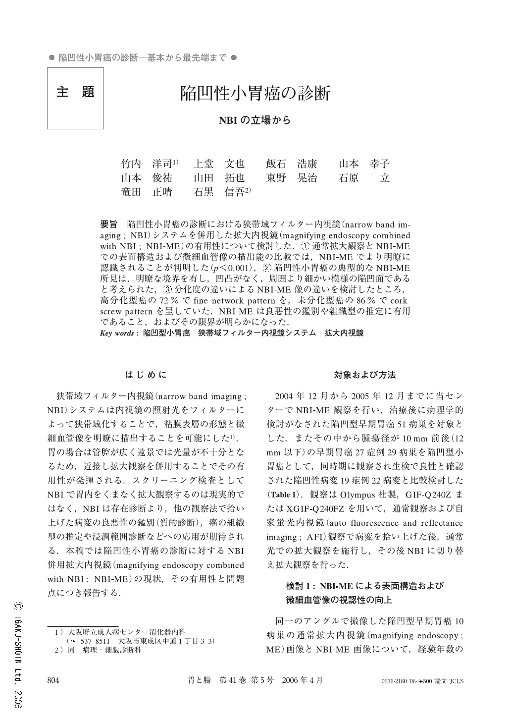
要旨 陥凹性小胃癌の診断における狭帯域フィルター内視鏡(narrow band imaging ; NBI)システムを併用した拡大内視鏡(magnifying endoscopy combined with NBI ; NBI-ME)の有用性について検討した.①通常拡大観察とNBI-MEでの表面構造および微細血管像の描出能の比較では,NBI-MEでより明瞭に認識されることが判明した(p<0.001),②陥凹性小胃癌の典型的なNBI-ME所見は,明瞭な境界を有し,凹凸がなく,周囲より細かい模様の陥凹面であると考えられた,③分化度の違いによるNBI-ME像の違いを検討したところ,高分化型癌の72%でfine network patternを,未分化型癌の86%でcorkscrew patternを呈していた.NBI-MEは良悪性の鑑別や組織型の推定に有用であること,およびその限界が明らかになった.
We evaluated the efficacy of magnifying endoscopy combined with narrow band imaging (NBI-ME) for small depressed type gastric cancer. ① We compared the visibility of NBI-ME and ordinary magnifying endoscopy (ME) for structure and vascular pattern of depressed-type gastric cancer. They could be recognized more clearly with NBI-ME image (p<0.001). ② We investigated the typical NBI-ME findings of small depressed-type gastric cancer. They were the presence of a demarcation line, disappearance of surface structure and difference in gland size and pattern compared with the surrounding mucosa. ③ The NBI-ME findings of gastric cancer correlated well with the histological differentiation of gastric cancer. The fine network pattern was seen in well differentiated adenocarcinoma (72%) and the corkscrew pattern in undifferentiated adenocarcinoma (86%). We reported the efficacy and limits of NBI-ME for making a diagnosis of carcinoma and an estimation of differentiation from gastric cancer.

Copyright © 2006, Igaku-Shoin Ltd. All rights reserved.


