Japanese
English
- 有料閲覧
PPV購入案内
1,320円 (1,200円+税10%) こちらは、96ページから100ページの文献です。購入には医書.jp本会員登録が必要です。
会員登録完了後に改めて上記「購入サイトへ」を選択してください。
または「購入サイトへ」選択後に「購入ログイン」、「新規会員登録」の順に進んでください。
会員登録について詳しくはをご確認ください。
- Abstract 文献概要
- 1ページ目 Look Inside
- 参考文献 Reference
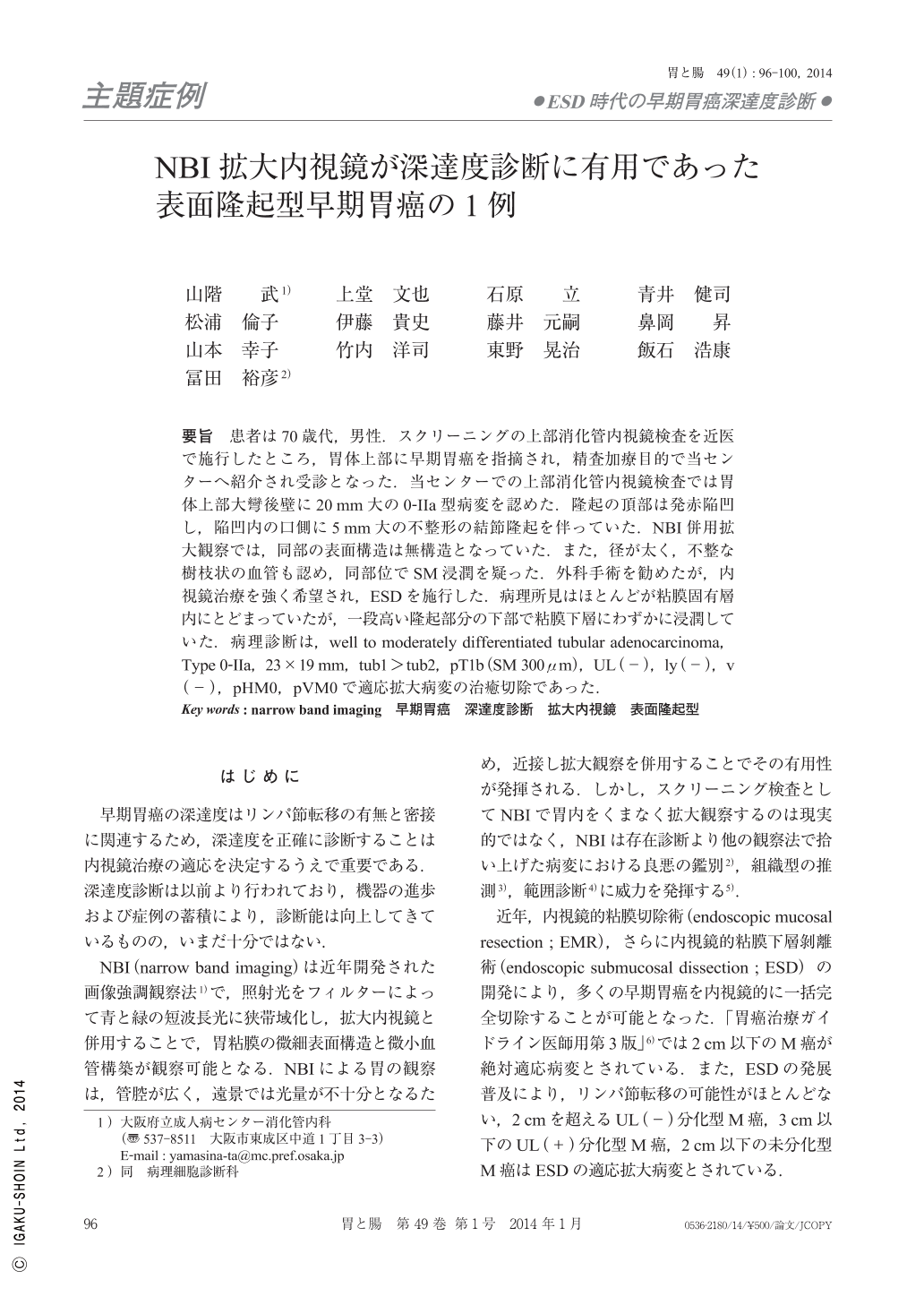
要旨 患者は70歳代,男性.スクリーニングの上部消化管内視鏡検査を近医で施行したところ,胃体上部に早期胃癌を指摘され,精査加療目的で当センターへ紹介され受診となった.当センターでの上部消化管内視鏡検査では胃体上部大彎後壁に20mm大の0-IIa型病変を認めた.隆起の頂部は発赤陥凹し,陥凹内の口側に5mm大の不整形の結節隆起を伴っていた.NBI併用拡大観察では,同部の表面構造は無構造となっていた.また,径が太く,不整な樹枝状の血管も認め,同部位でSM浸潤を疑った.外科手術を勧めたが,内視鏡治療を強く希望され,ESDを施行した.病理所見はほとんどが粘膜固有層内にとどまっていたが,一段高い隆起部分の下部で粘膜下層にわずかに浸潤していた.病理診断は,well to moderately differentiated tubular adenocarcinoma,Type 0-IIa,23×19mm,tub1>tub2,pT1b(SM 300μm),UL(-),ly(-),v(-),pHM0,pVM0で適応拡大病変の治癒切除であった.
A man in his 70's was referred to our hospital for endoscopic treatment of an early gastric cancer. Esophagogastroduodenoscopy showed a 0-IIa type superficial cancer 20mm in size at the greater curvature of the upper gastric body. The lesion had an irregular nodule, which was 5mm in size, in the proximal part of the tumor. Magnifying narrow band imaging endoscopy revealed irregular microvessels and the absence of microstructure pattern in the proximal nodule. We assumed there was submucosal invasion of the cancer at the nodule, therefore, recommended that he receive a surgical operation. Despite our strong recommendation, he insisted on undergoing ESD(endoscopic submucosal dissection). After obtaining written informed consent for the anticipated result, possible risk of complications and additional surgical resection in case of non-curative resection, ESD was undertaken for the lesion. Pathological diagnosis of the resected specimen was moderately differentiated tubular adenocarcinoma, Type 0-IIa, 23×19mm, pT1b(SM 300μm), UL(-), ly(-), v(-), pHM0, pVM0 ; and it fulfilled curative resection criteria for an expanded-indication lesion.
Copyright © 2014, Igaku-Shoin Ltd. All rights reserved.