Japanese
English
- 有料閲覧
- Abstract 文献概要
- 1ページ目 Look Inside
- 参考文献 Reference
- サイト内被引用 Cited by
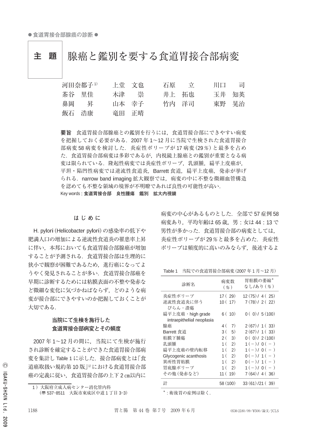
要旨 食道胃接合部腺癌との鑑別を行うには,食道胃接合部にできやすい病変を把握しておく必要がある.2007年1~12月に当院で生検された食道胃接合部病変58病変を検討した.炎症性ポリープが17病変(29%)と最多を占めた.食道胃接合部病変は多彩であるが,内視鏡上腺癌との鑑別が重要となる病変は限られている.隆起性病変では炎症性ポリープ,乳頭腫,扁平上皮癌が,平坦・陥凹性病変では逆流性食道炎,Barrett食道,扁平上皮癌,発赤が挙げられる.narrow band imaging拡大観察では,病変の中に不整な微細血管構造を認めても不整な領域の境界が不明瞭であれば良性の可能性が高い.
To diagnose esophagogastric adenocarcinoma, the variety and frequency of esophagogastric lesions including benign tumors should be discerned. We analyzed 58 histologically diagnosed esophagogastric lesions observed endoscopically from Jan. 2007 to Dec. 2007. The most frequently observed lesions were inflammatory polyps(17 lesions,29%).
There are varieties of esophagogastric lesions, but, the lesions which need differential diagnosis are limited. For example, for the protruding lesions, inflammatory polyps, papilloma and squamous cell carcinoma are important. For the flat or depressed lesions, reflex esophagitis,Barrett esophagus, squamous cell carcinoma and reddishness are important. Irregular micro-vessels observed by magnified endoscopy combined with narrow band imaging(NBI-ME)indicate the lesion might be malignant. However, the unclear margin of the irregular micro-vessel area rather suggests the lesion might be benign.

Copyright © 2009, Igaku-Shoin Ltd. All rights reserved.


