Japanese
English
- 有料閲覧
- Abstract 文献概要
- 1ページ目 Look Inside
- 参考文献 Reference
- サイト内被引用 Cited by
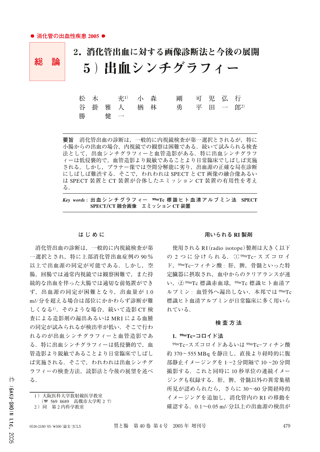
要旨 消化管出血の診断は,一般的に内視鏡検査が第一選択とされるが,特に小腸からの出血の場合,内視鏡での観察は困難である.続いて試みられる検査法として,出血シンチグラフィーと血管造影がある.特に出血シンチグラフィーは低侵襲的で,血管造影より鋭敏であることより日常臨床でしばしば実施される.しかし,プラナー像では空間分解能に劣り,出血源の正確な局在診断にしばしば難渋する.そこで,われわれはSPECTとCT画像の融合像あるいはSPECT装置とCT装置が合体したエミッションCT装置の有用性を考える.
For diagnosis and localization of gastrointestinal (GI) bleeding, GI endoscopy is commonly performed as a first step. However, bleeding in the small bowel cannot be directly visualized by endoscopy. In such a case, angiography or nuclear scintigraphy is performed. Nuclear scintigraphy is a noninvasive method for diagnosing accurately GI bleeding, especially in the small bowel. However, the planar image is poor in spatial resolution, therefore it is difficult to localize the bleeding site exactly. We consider that the fusion of CT and SPECT images or an emission CT machine consisting of CT and SPECT machines may be useful for the localization of GI bleeding.

Copyright © 2005, Igaku-Shoin Ltd. All rights reserved.


