Japanese
English
- 有料閲覧
PPV購入案内
2,750円 (2,500円+税10%) こちらは、1031ページから1038ページの文献です。購入には医書.jp本会員登録が必要です。
会員登録完了後に改めて上記「購入サイトへ」を選択してください。
または「購入サイトへ」選択後に「購入ログイン」、「新規会員登録」の順に進んでください。
会員登録について詳しくはをご確認ください。
- Abstract 文献概要
- 1ページ目 Look Inside
- 参考文献 Reference
要 旨
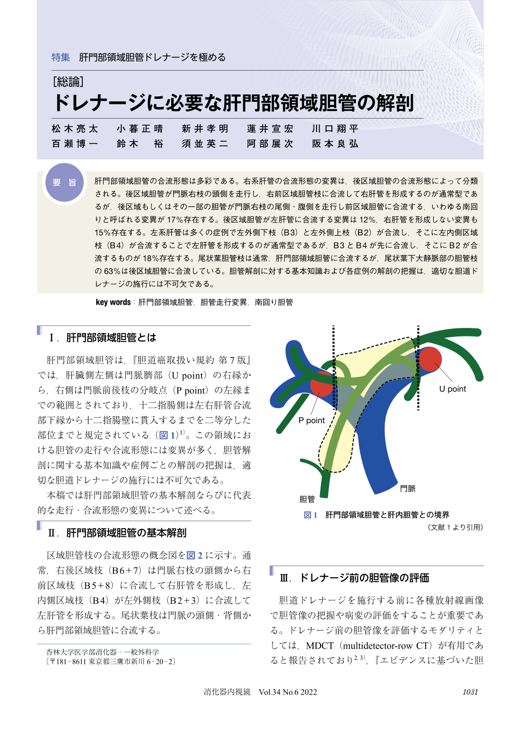
肝門部領域胆管の合流形態は多彩である。右系肝管の合流形態の変異は,後区域胆管の合流形態によって分類される。後区域胆管が門脈右枝の頭側を走行し,右前区域胆管枝に合流して右肝管を形成するのが通常型であるが,後区域もしくはその一部の胆管が門脈右枝の尾側・腹側を走行し前区域胆管に合流する,いわゆる南回りと呼ばれる変異が17%存在する。後区域胆管が左肝管に合流する変異は12%,右肝管を形成しない変異も15%存在する。左系肝管は多くの症例で左外側下枝(B3)と左外側上枝(B2)が合流し,そこに左内側区域枝(B4)が合流することで左肝管を形成するのが通常型であるが,B3とB4が先に合流し,そこにB2が合流するものが18%存在する。尾状葉胆管枝は通常,肝門部領域胆管に合流するが,尾状葉下大静脈部の胆管枝の63%は後区域胆管に合流している。胆管解剖に対する基本知識および各症例の解剖の把握は,適切な胆道ドレナージの施行には不可欠である。
The perihilar bile duct has a variety of confluence pattern. Variation in the confluence of the right hepatic bile duct is mainly determined by the confluence pattern of the posterior bile duct. In general, the posterior bile duct joins the anterior bile duct via supraportal route to form the right hepatic bile duct. But the posterior bile duct joins the anterior bile duct via infraportal route in 17% of cases. The posterior bile duct joins the left hepatic duct in 12% of cases, thus the right hepatic duct is absent in 15% of cases. The left hepatic bile duct is generally formed by the confluence of B4 and B2+3. But the left hepatic bile duct formed by the confluence of B2 and B3+4 in 18% of cases. The biliary branches of the caudate lobe generally join the perihilar bile duct. Of these, the biliary branches in the paracaval portion joins the posterior bile duct in 63% of cases. Basic knowledge and individual variation of the anatomy of the perihilar bile duct are essential for adequate biliary drainage.
© tokyo-igakusha.co.jp. All right reserved.