Japanese
English
- 有料閲覧
PPV購入案内
1,650円 (1,500円+税10%) こちらは、475ページから479ページの文献です。購入には医書.jp本会員登録が必要です。
会員登録完了後に改めて上記「購入サイトへ」を選択してください。
または「購入サイトへ」選択後に「購入ログイン」、「新規会員登録」の順に進んでください。
会員登録について詳しくはをご確認ください。
- Abstract 文献概要
- 1ページ目 Look Inside
- 参考文献 Reference
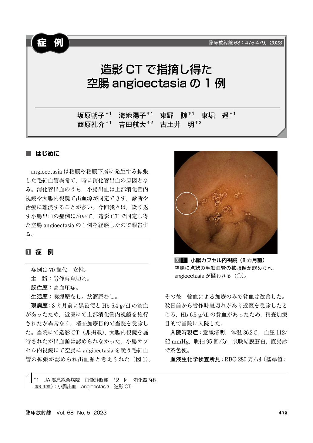
angioectasiaは粘膜や粘膜下層に発生する拡張した毛細血管異常で,時に消化管出血の原因となる。消化管出血のうち,小腸出血は上部消化管内視鏡や大腸内視鏡で出血源が同定できず,診断や治療に難渋することが多い。今回我々は,繰り返す小腸出血の症例において,造影CTで同定し得た空腸angioectasiaの1例を経験したので報告する。
The patient was a woman in her seventies. Eight months ago, she had experienced hematochezia and was found to have anemia with a hemoglobin of 5.4 g/dl. Esophagogastroduodenoscopy and colonoscopy could not identify the source of bleeding. Capsule endoscopy revealed jejunal angioectasia suspected to be the cause. She was transfused and her anemia improved. This time, she complained shortness of breath on exertion, and because she was found to have anemia with a hemoglobin of 6.5 g/dl, she was hospitalized. Contrast enhanced CT demonstrated focal punctate areas and bulbous swelling of the intramural vessels in the wall of the jejunum on enteric phase(50 seconds after the beginning of injection). These findings consisted with angioectasia. Enteric phase is useful for detection of angioectasia.
Copyright © 2023, KANEHARA SHUPPAN Co.LTD. All rights reserved.