Japanese
English
- 有料閲覧
- Abstract 文献概要
- 1ページ目 Look Inside
- 参考文献 Reference
- サイト内被引用 Cited by
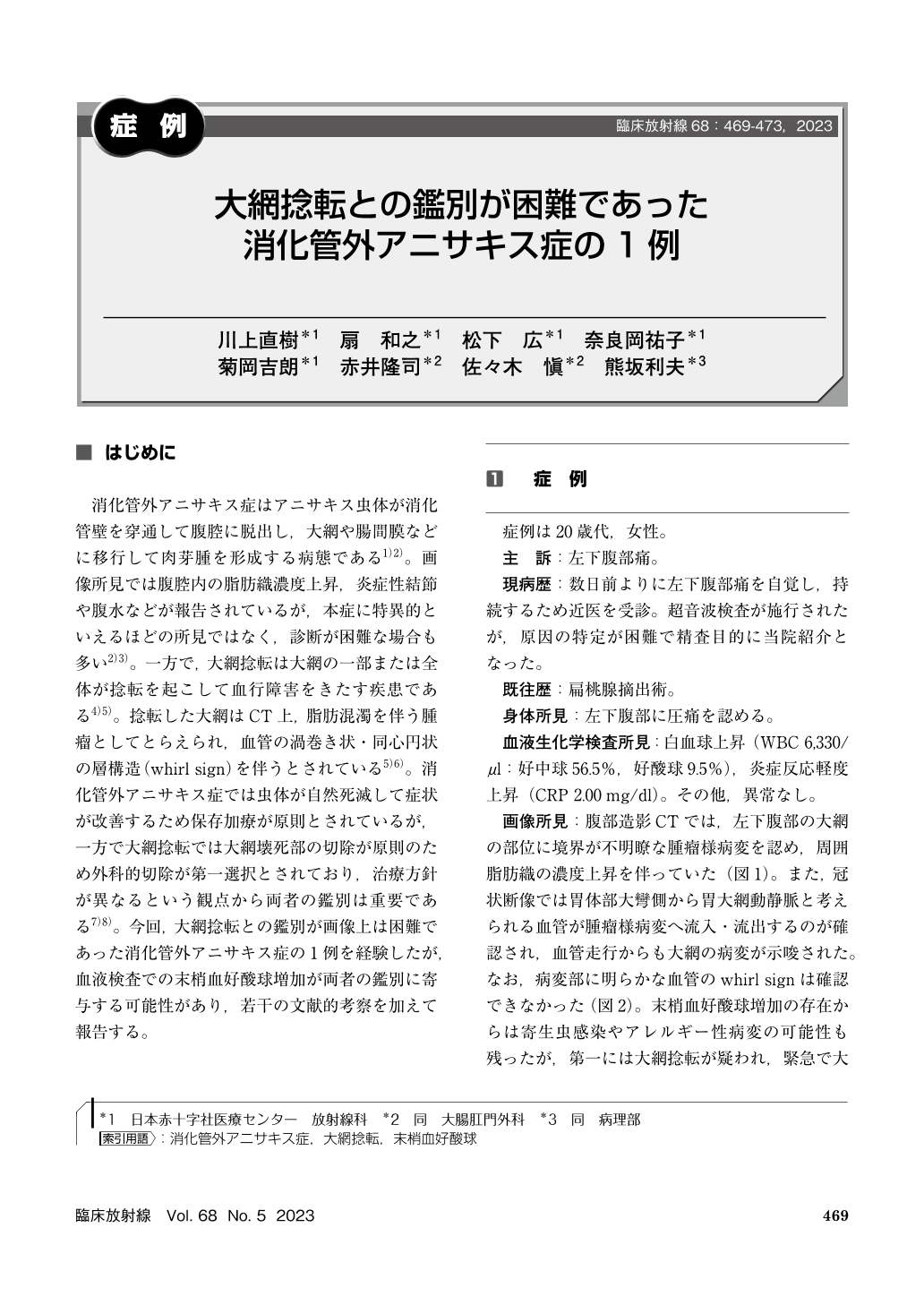
消化管外アニサキス症はアニサキス虫体が消化管壁を穿通して腹腔に脱出し,大網や腸間膜などに移行して肉芽腫を形成する病態である1)2)。画像所見では腹腔内の脂肪織濃度上昇,炎症性結節や腹水などが報告されているが,本症に特異的といえるほどの所見ではなく,診断が困難な場合も多い2)3)。一方で,大網捻転は大網の一部または全体が捻転を起こして血行障害をきたす疾患である4)5)。捻転した大網はCT上,脂肪混濁を伴う腫瘤としてとらえられ,血管の渦巻き状・同心円状の層構造(whirl sign)を伴うとされている5)6)。消化管外アニサキス症では虫体が自然死滅して症状が改善するため保存加療が原則とされているが,一方で大網捻転では大網壊死部の切除が原則のため外科的切除が第一選択とされており,治療方針が異なるという観点から両者の鑑別は重要である7)8)。今回,大網捻転との鑑別が画像上は困難であった消化管外アニサキス症の1例を経験したが,血液検査での末梢血好酸球増加が両者の鑑別に寄与する可能性があり,若干の文献的考察を加えて報告する。
Extra-gastrointestinal anisakiasis is a rare disorder characterized by penetration of live larvae through gastrointestinal wall into peritoneal cavity, such as omentum and mesentery. A 25-year-old woman who complained of left lower abdominal pain visited our hospital for further evaluation. Contrast-enhanced CT showed a mass-like lesion in the left omentum, with increased attenuation of surrounding fatty tissues.
Omental torsion was suspected, and partial omentectomy was performed. Postoperative pathology revealed anisakiasis larvae, leading to a definitive diagnosis of extra-gastrointestinal anisakiasis. Extra-gastrointestinal anisakiasis has many similarities with omental torsion on CT findings, leading to difficulties to differentiate these two disorders. Eosinophils elevation in blood tests may contribute to the correct diagnosis, along with detailed taking of food history. Although rare incidence, extra-gastrointestinal anisakiasis should be considered for the differential diagnosis of painful peritoneal masses, when eosinophils elevation and the history of raw fish ingestion are present.

Copyright © 2023, KANEHARA SHUPPAN Co.LTD. All rights reserved.


