Japanese
English
- 有料閲覧
PPV購入案内
1,650円 (1,500円+税10%) こちらは、643ページから656ページの文献です。購入には医書.jp本会員登録が必要です。
会員登録完了後に改めて上記「購入サイトへ」を選択してください。
または「購入サイトへ」選択後に「購入ログイン」、「新規会員登録」の順に進んでください。
会員登録について詳しくはをご確認ください。
- Abstract 文献概要
- 1ページ目 Look Inside
- 参考文献 Reference
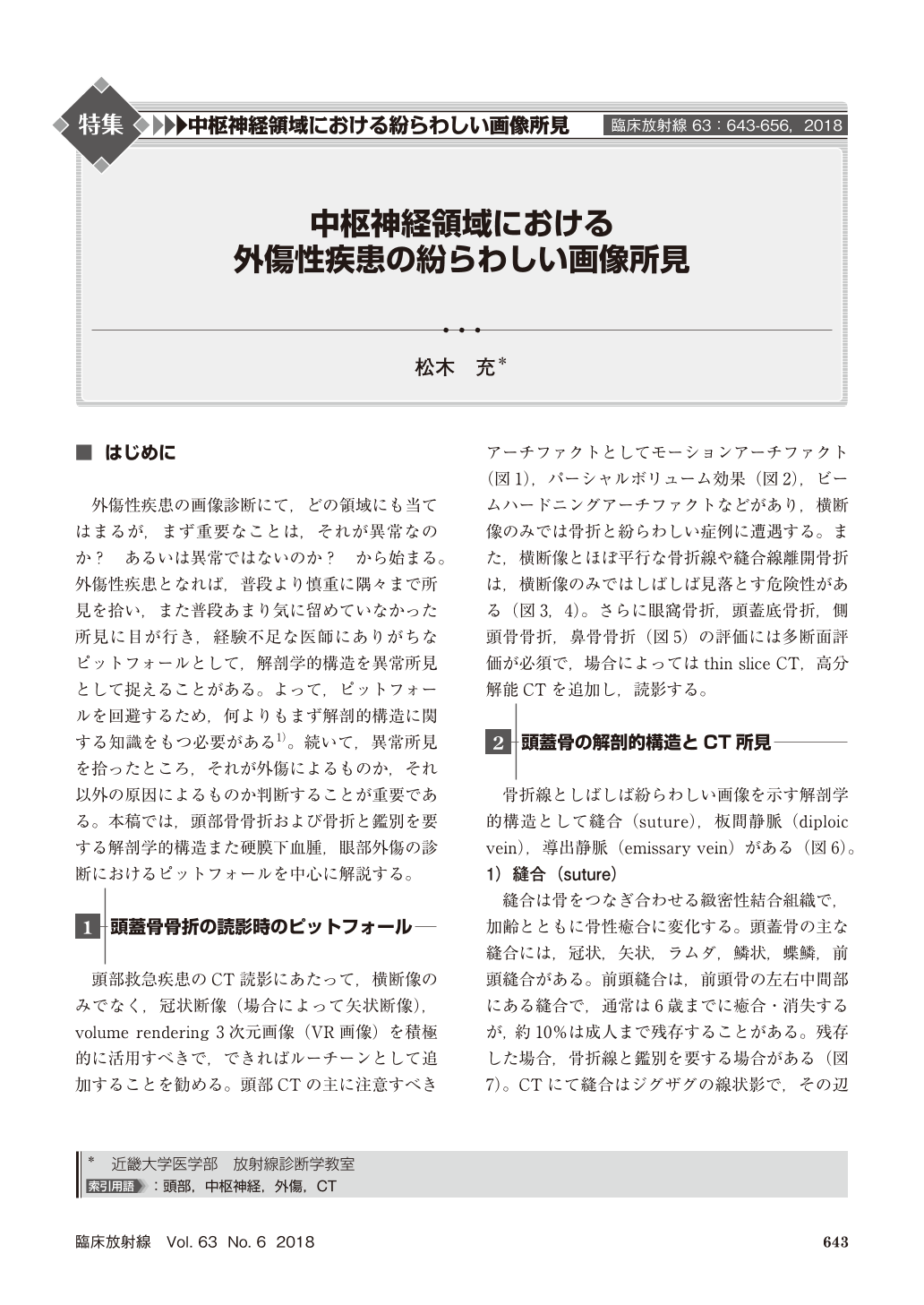
外傷性疾患の画像診断にて,どの領域にも当てはまるが,まず重要なことは,それが異常なのか? あるいは異常ではないのか? から始まる。外傷性疾患となれば,普段より慎重に隅々まで所見を拾い,また普段あまり気に留めていなかった所見に目が行き,経験不足な医師にありがちなピットフォールとして,解剖学的構造を異常所見として捉えることがある。よって,ピットフォールを回避するため,何よりもまず解剖的構造に関する知識をもつ必要がある1)。続いて,異常所見を拾ったところ,それが外傷によるものか,それ以外の原因によるものか判断することが重要である。本稿では,頭部骨骨折および骨折と鑑別を要する解剖学的構造また硬膜下血腫,眼部外傷の診断におけるピットフォールを中心に解説する。
In imaging diagnosis of traumatic diseases in any organ, it is important to decide it to be normal or abnormal. In imaging diagnosis of traumatic diseases, we observe images more carefully than usual, and interpret in more detail than usual. Then, doctors lacking experience tend to interpret normal variations as abnormal findings in organ. Therefore, in order to avoid the pitfalls, it is necessary to first have knowledge about the anatomical structure. Subsequently, when we pick up abnormal findings, it is important to decide whether it is due to trauma or other causes. In this paper, we introduce pitfalls in imaging diagnosis of head bone fracture, subdural hematoma and ocular trauma.
Copyright © 2018, KANEHARA SHUPPAN Co.LTD. All rights reserved.