Japanese
English
- 有料閲覧
PPV購入案内
1,650円 (1,500円+税10%) こちらは、471ページから474ページの文献です。購入には医書.jp本会員登録が必要です。
会員登録完了後に改めて上記「購入サイトへ」を選択してください。
または「購入サイトへ」選択後に「購入ログイン」、「新規会員登録」の順に進んでください。
会員登録について詳しくはをご確認ください。
- Abstract 文献概要
- 1ページ目 Look Inside
- 参考文献 Reference
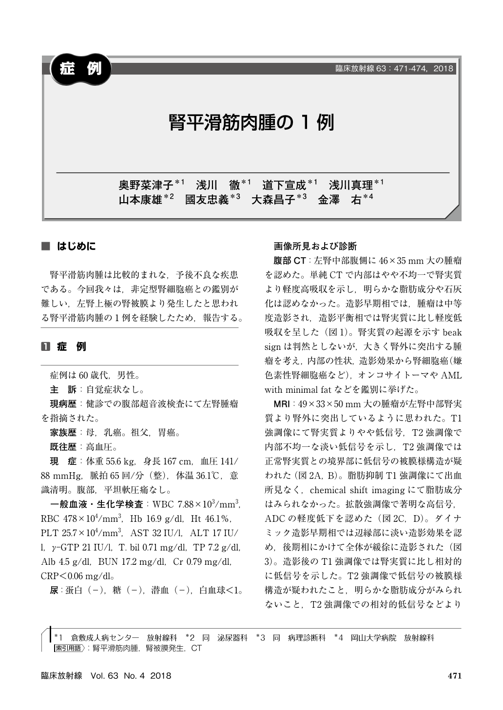
腎平滑筋肉腫は比較的まれな,予後不良な疾患である。今回我々は,非定型腎細胞癌との鑑別が難しい,左腎上極の腎被膜より発生したと思われる腎平滑筋肉腫の1例を経験したため,報告する。
Non-contrast CT in a male in his 60s showed a 50-mm solid tumor with slight internal inhomogeneity and slightly higher absorbance than renal parenchyma on the left renal ventral side. Moderate contrast was obtained in early-phase contrast CT, with slightly lower absorbance than renal parenchyma in the equilibrium phase. T2WI MRI showed slightly lower signals with internal inhomogeneity, no fat components, and high-intensity diffusion-weighted signals. Laparoscopic left renal partial resection was performed for suspected papillary renal cell carcinoma. Histopathologically, the tumor had developed on the renal capsule, and leiomyosarcoma was diagnosed from immunostaining. This case illustrates that leiomyosarcoma should be considered for a solid tumor on the renal capsule.
Copyright © 2018, KANEHARA SHUPPAN Co.LTD. All rights reserved.