Japanese
English
- 有料閲覧
PPV購入案内
1,320円 (1,200円+税10%) こちらは、201ページから205ページの文献です。購入には医書.jp本会員登録が必要です。
会員登録完了後に改めて上記「購入サイトへ」を選択してください。
または「購入サイトへ」選択後に「購入ログイン」、「新規会員登録」の順に進んでください。
会員登録について詳しくはをご確認ください。
- Abstract 文献概要
- 1ページ目 Look Inside
- 参考文献 Reference
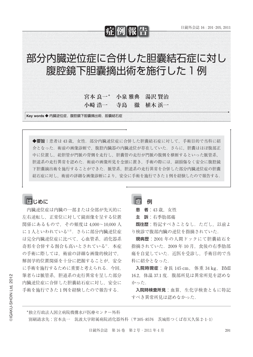
◆要旨:患者は43歳,女性.部分内臓逆位症に合併した胆囊結石症に対して,手術目的で当科に紹介となった.術前の画像診断で,腹腔内臓器の内臓逆位が存在していた.さらに,胆囊はほぼ腹部正中に位置し,総胆管が門脈の背側を走行し,胆囊管の走行が門脈の腹側を横断するといった脈管系,胆道系の走行異常を認めた.術前の画像所見を念頭に置き,手術の際には,副損傷なく安全に腹腔鏡下胆囊摘出術を施行することができた.脈管系,胆道系の走行異常を合併した部分内臓逆位症の胆囊結石症に対し,術前の詳細な画像診断により,安全に手術を施行できた1例を経験したので報告する.
A 40-year-old female patient was referred to us for surgery of gallstones accompanied by partial situs inversus. Preoperative diagnostic imaging revealed heterotaxia of the abdominal organs. At the hepatic hilus, abnormal arrangements of the vascular and biliary systems were noted, such as the gallbladder located approximately in the median abdominal position, the common bile duct arranged dorsal to the portal vein, and the cystic duct crossing the ventral side of the portal vein. With these findings from preoperative imaging taken into account, laparoscopic cholecystectomy was carried out safely without causing any additional injury. We report a case of gallstones accompanied by partial situs inversus(abnormal arrangements of the vascular and biliary systems)where surgery could be performed safely after detailed preoperative diagnostic imaging.
Copyright © 2011, JAPAN SOCIETY FOR ENDOSCOPIC SURGERY All rights reserved.