Japanese
English
- 有料閲覧
PPV購入案内
1,320円 (1,200円+税10%) こちらは、99ページから103ページの文献です。購入には医書.jp本会員登録が必要です。
会員登録完了後に改めて上記「購入サイトへ」を選択してください。
または「購入サイトへ」選択後に「購入ログイン」、「新規会員登録」の順に進んでください。
会員登録について詳しくはをご確認ください。
- Abstract 文献概要
- 1ページ目 Look Inside
- 参考文献 Reference
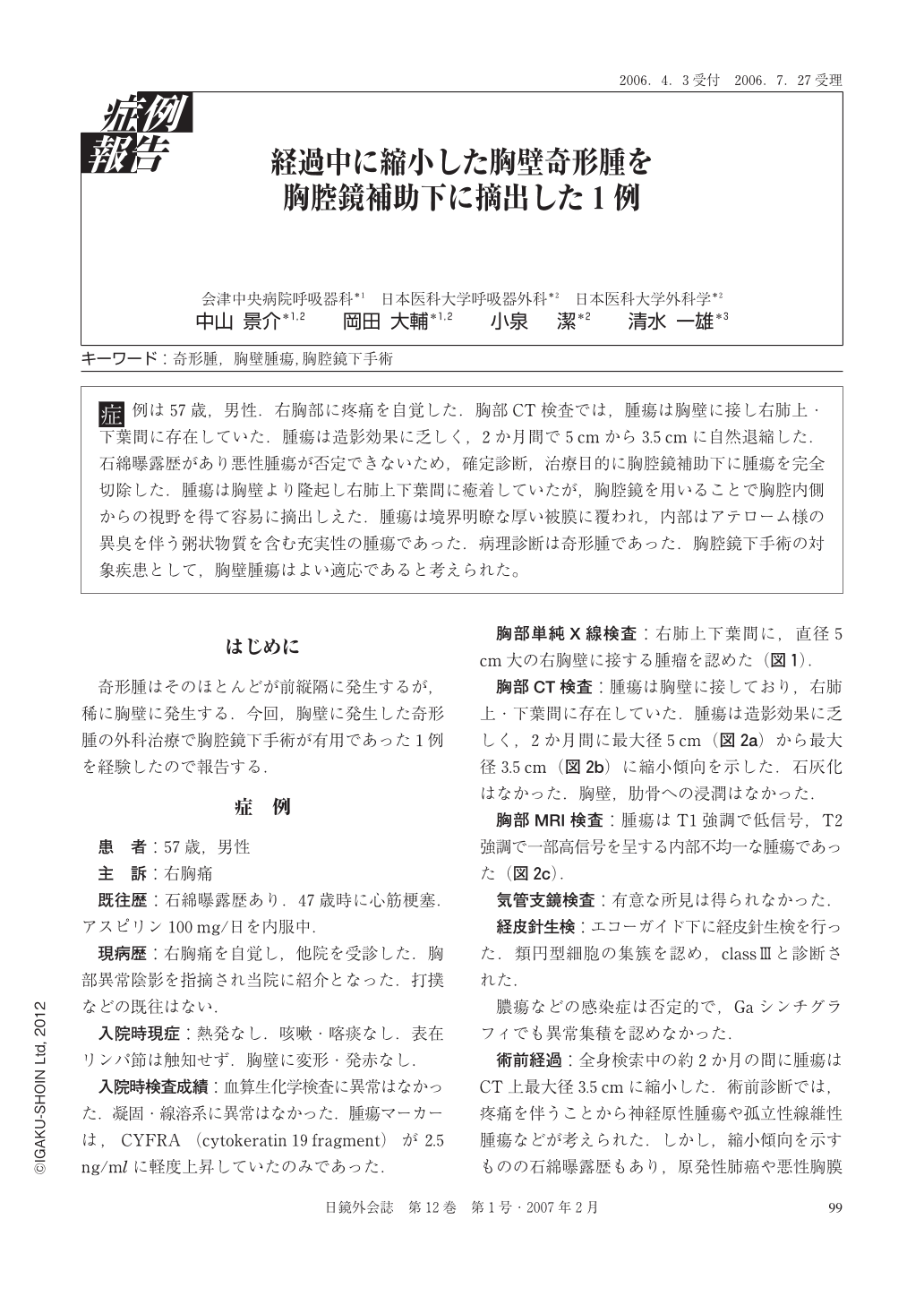
症例は57歳,男性.右胸部に疼痛を自覚した.胸部CT検査では,腫瘍は胸壁に接し右肺上・下葉間に存在していた.腫瘍は造影効果に乏しく,2か月間で5cmから3.5cmに自然退縮した.石綿曝露歴があり悪性腫瘍が否定できないため,確定診断,治療目的に胸腔鏡補助下に腫瘍を完全切除した.腫瘍は胸壁より隆起し右肺上下葉間に癒着していたが,胸腔鏡を用いることで胸腔内側からの視野を得て容易に摘出しえた.腫瘍は境界明瞭な厚い被膜に覆われ,内部はアテローム様の異臭を伴う粥状物質を含む充実性の腫瘍であった.病理診断は奇形腫であった.胸腔鏡下手術の対象疾患として,胸壁腫瘍はよい適応であると考えられた。
A 57-year-old man experienced a pain in the right chest. Computed tomography of the chest showed a tumor between the upper and lower lobes of the right lung, which was in contact with the chest wall. The tumor with less contrast enhancement spontaneously decreased from 5 to 3.5 cm in two months. Malignancy could not be ruled out because of his previous exposure to asbestos, and thus the tumor was completely resected thoracoscopically for definite diagnosis and treatment. Although the tumor protruded from the chest wall and adhered to the region between the upper and lower lobes of the right lung, a thoracoscope allowed easy resection of the tumor with view from inside the thoracic cavity. Surrounded by a well-demarcated thick capsule, the tumor was solid and contained atheromatous, pultaceous material with an unpleasant odor. Pathological diagnosis was teratoma. Chest wall tumors appear to be a good indication for thoracoscopic surgery.
Copyright © 2007, JAPAN SOCIETY FOR ENDOSCOPIC SURGERY All rights reserved.