Japanese
English
- 有料閲覧
PPV購入案内
2,200円 (2,000円+税10%) こちらは、875ページから882ページの文献です。購入には医書.jp本会員登録が必要です。
会員登録完了後に改めて上記「購入サイトへ」を選択してください。
または「購入サイトへ」選択後に「購入ログイン」、「新規会員登録」の順に進んでください。
会員登録について詳しくはをご確認ください。
- Abstract 文献概要
- 1ページ目 Look Inside
- 参考文献 Reference
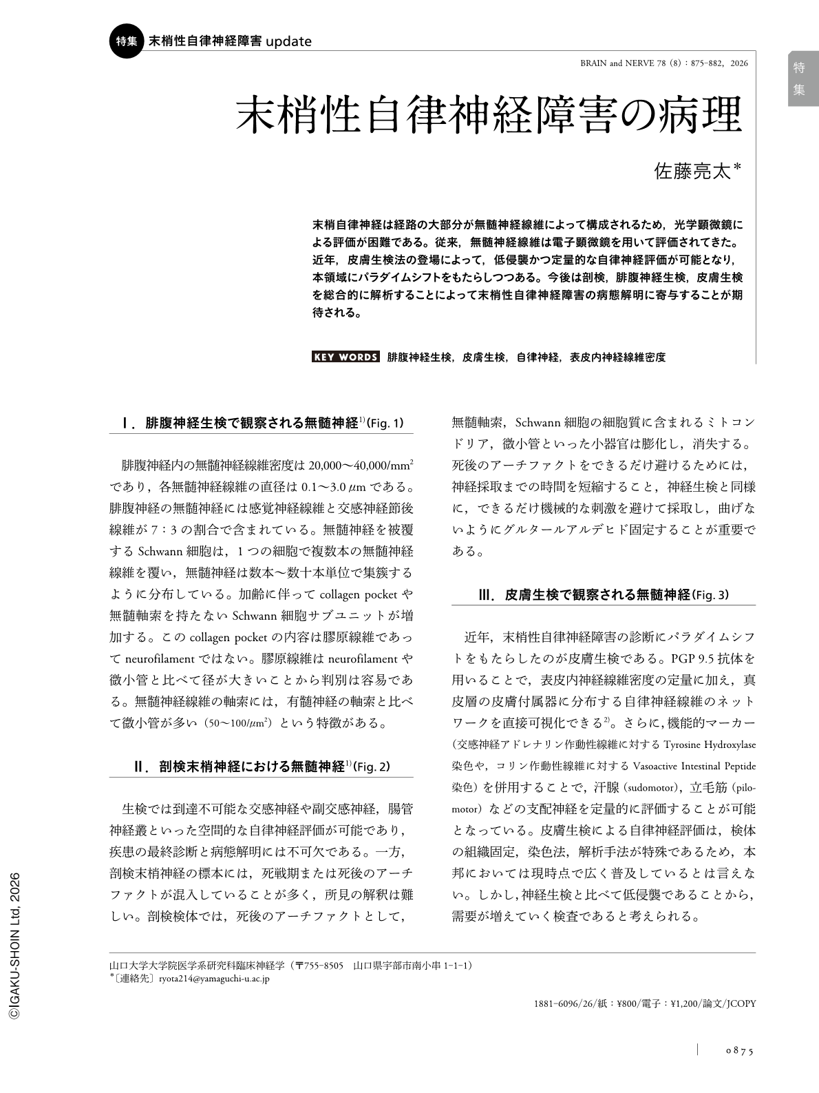
末梢自律神経は経路の大部分が無髄神経線維によって構成されるため,光学顕微鏡による評価が困難である。従来,無髄神経線維は電子顕微鏡を用いて評価されてきた。近年,皮膚生検法の登場によって,低侵襲かつ定量的な自律神経評価が可能となり,本領域にパラダイムシフトをもたらしつつある。今後は剖検,腓腹神経生検,皮膚生検を総合的に解析することによって末梢性自律神経障害の病態解明に寄与することが期待される。
Abstract
The peripheral autonomic nervous system predominantly comprises unmyelinated fibers, which are difficult to evaluate using conventional light microscopy. Historically, morphological assessment of unmyelinated fibers relied on the electron microscopy of sural nerve biopsy specimens, which is a technically demanding approach with limited accessibility to autonomic structures. Skin biopsy with immunohistochemical staining for the pan-axonal marker PGP 9.5 has enabled minimally invasive and quantitative evaluation of somatic small (intraepidermal nerve fiber density) and autonomic fibers innervating sweat glands, arrector pili muscles, and dermal vasculature. An integrated analysis combining autopsy findings, sural nerve biopsy, and skin biopsy may clarify the pathophysiology of peripheral autonomic neuropathy.
Copyright © 2026, Igaku-Shoin Ltd. All rights reserved.