Japanese
English
- 有料閲覧
PPV購入案内
1,320円 (1,200円+税10%) こちらは、267ページから274ページの文献です。購入には医書.jp本会員登録が必要です。
会員登録完了後に改めて上記「購入サイトへ」を選択してください。
または「購入サイトへ」選択後に「購入ログイン」、「新規会員登録」の順に進んでください。
会員登録について詳しくはをご確認ください。
- Abstract 文献概要
- 1ページ目 Look Inside
- 参考文献 Reference
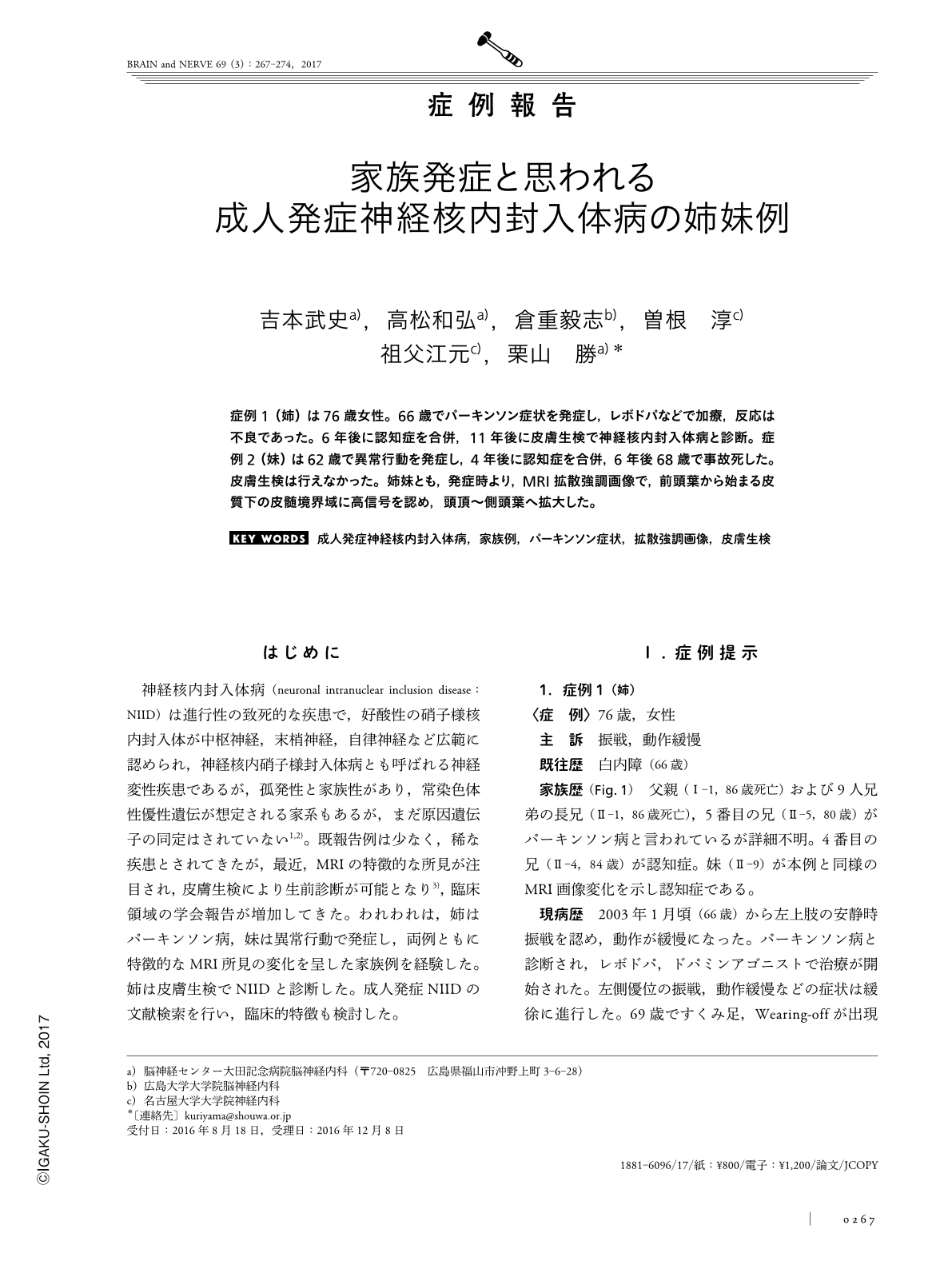
症例1(姉)は76歳女性。66歳でパーキンソン症状を発症し,レボドパなどで加療,反応は不良であった。6年後に認知症を合併,11年後に皮膚生検で神経核内封入体病と診断。症例2(妹)は62歳で異常行動を発症し,4年後に認知症を合併,6年後68歳で事故死した。皮膚生検は行えなかった。姉妹とも,発症時より,MRI拡散強調画像で,前頭葉から始まる皮質下の皮髄境界域に高信号を認め,頭頂〜側頭葉へ拡大した。
Abstract
Neuronal intranuclear inclusion disease (NIID) in two adult siblings (both women, aged 76 and 68 years) is reported on here. The elder sister had a resting tremor and bradykinesia at age 66 years, and treatment with L-DOPA was initiated(L-3, 4-dihydroxyphenylalanine). Three years later, she showed a frozen gait that was associated with the medication wearing off. The clinical manifestations did not improve with the administration of antiparkinson drugs. Six years later, she showed impaired cognitive functions, which had occured gradually, and she began to take donepezil. At age 76, she was diagnosed with NIID based on a skin biopsy. The younger sister exhibited peculiar behaviors at age 62 years, and showed impaired cognitive function 4 years later. At age 68 years, she died because of an accident in the bath tub. In both cases, diffusion-weighted magnetic resonance imaging (DWI) showed high-intensity signals in the U fiber area of the corticomedullary junction. These signals began in the frontal lobe at the initial stages of the disease, and extended to the parietal and temporal lobes at later stages. High-intensity signal areas were detected in the deep white matter in T2-weighted and fluid-attenuated inversion recovery (FLAIR) images in the elder sister. The histological examination via a skin biopsy was useful in diagnosing NIID.
(Received August 18, 2016; Accepted December 8, 2016; Published March 1, 2017)
Copyright © 2017, Igaku-Shoin Ltd. All rights reserved.