Japanese
English
- 有料閲覧
PPV購入案内
1,320円 (1,200円+税10%) こちらは、529ページから533ページの文献です。購入には医書.jp本会員登録が必要です。
会員登録完了後に改めて上記「購入サイトへ」を選択してください。
または「購入サイトへ」選択後に「購入ログイン」、「新規会員登録」の順に進んでください。
会員登録について詳しくはをご確認ください。
- Abstract 文献概要
- 1ページ目 Look Inside
I.緒言
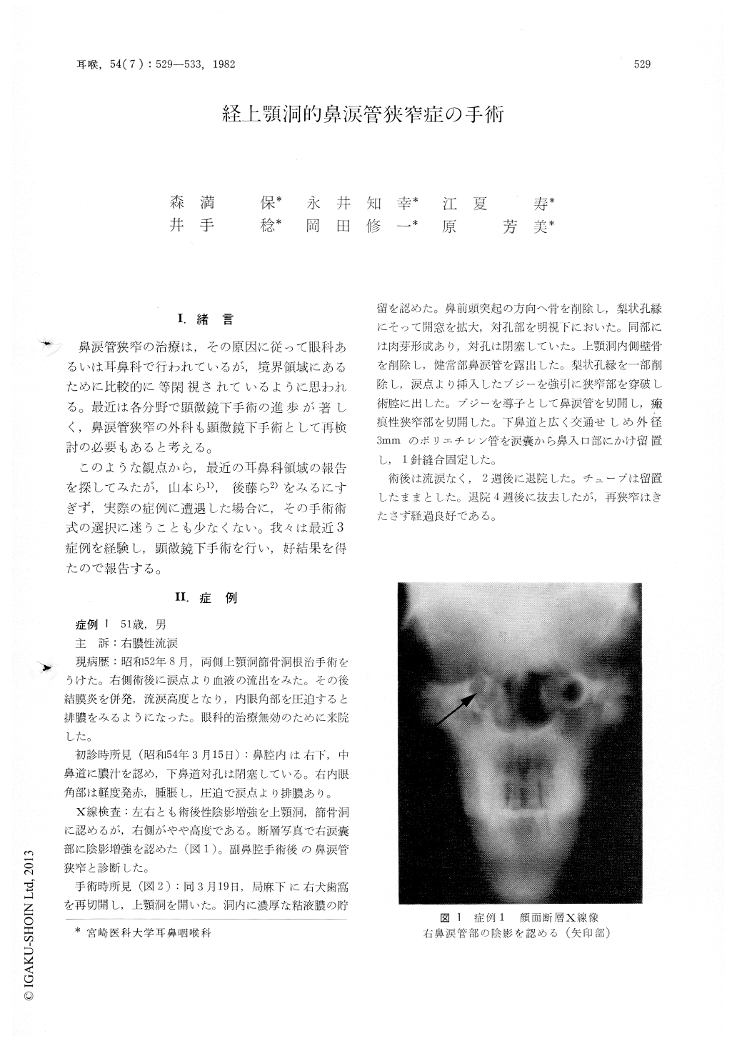
鼻涙管狭窄の治療は,その原因に従って眼科あるいは耳鼻科で行われているが,境界領域にあるために比較的に等閑視されているように思われる。最近は各分野で顕微鏡下手術の進歩が著しく,鼻涙管狭窄の外科も顕微鏡下手術として再検討の必要もあると考える。
このような観点から,最近の耳鼻科領域の報告を探してみたが,山本ら1),後藤ら2)をみるにすぎず,実際の症例に遭遇した場合に,その手術術式の選択に迷うことも少なくない。我々は最近3症例を経験し,顕微鏡下手術を行い,好結果を得たので報告する。
Three cases of the stenotic nasolacrimal duct treated surgically with transmaxillary approach under an operating microscope were reported. The causes of the stenosis were cicatrization at the nasal orifice after sinus operation, dacryocystitis and dacryocyst mucocele respectively.
The surgical procedures were as follows: 1) Open the maxillary sinus from the canine fossa. 2) Removal of the medial bony wall of the sinus and exposure of the nasolacrimal duct. 3) Open the nasal orifice of the duct by incisionof the mucous membrane of the inferior nasal meatus. 4) Bouginage of the lacrimal duct through the stenotic portion. 5) Removal or blunt dilatation of the stenosis. 6) Insertion of a polyethylene tube of 2-3 mm diameter to the lacrimal sack. In our cases, the inserted tubes were retaind in their place for one month in one case and for over one year in two cases with good result.
Copyright © 1982, Igaku-Shoin Ltd. All rights reserved.