Japanese
English
--------------------
耳下腺腫瘍の造影断層撮影
THE USE OF TOMOGRAPHY IN THE DIAGNOSIS OF PAROTID TUMORS
木村 淑志
1
Yoshiyuki Kimura
1
1順天堂大学医学部耳鼻咽喉科学教室
pp.13-17
発行日 1975年1月20日
Published Date 1975/1/20
DOI https://doi.org/10.11477/mf.1492208154
- 有料閲覧
PPV購入案内
1,320円 (1,200円+税10%) こちらは、13ページから17ページの文献です。
PPV(ペイ・パー・ビュー)とは、論文(記事)単位で購入・閲覧ができるサービスです。
購入には医書.jp本会員登録が必要です。
会員登録完了後に改めて上記「購入サイトへ」を選択してください。
または「購入サイトへ」選択後に「購入ログイン」、「新規会員登録」の順に進んでください。
会員登録について詳しくはをご確認ください。
購入には医書.jp本会員登録が必要です。
会員登録完了後に改めて上記「購入サイトへ」を選択してください。
または「購入サイトへ」選択後に「購入ログイン」、「新規会員登録」の順に進んでください。
会員登録について詳しくはをご確認ください。
- Abstract 文献概要
- 1ページ目 Look Inside
I.緒言
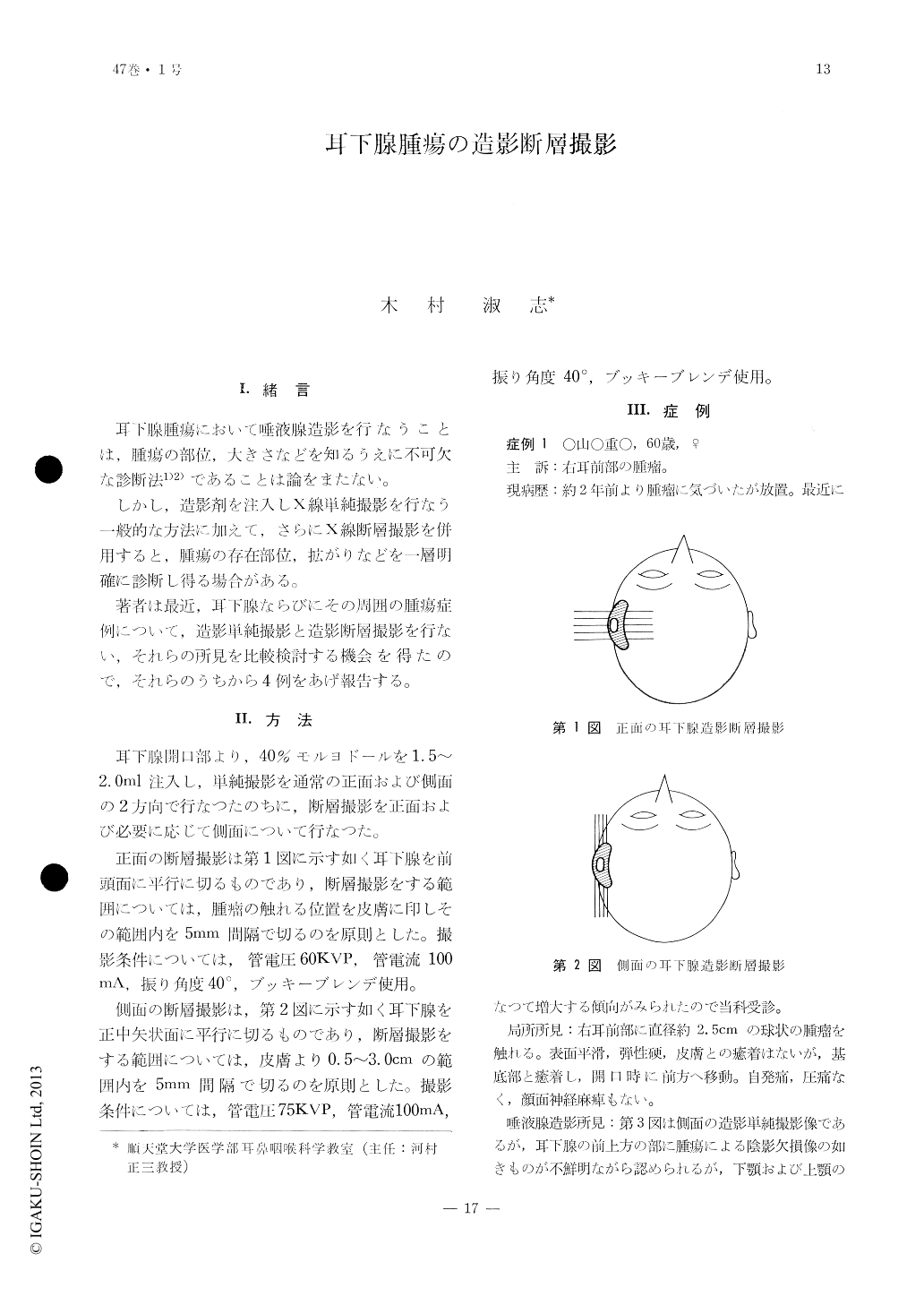
耳下腺腫瘍において唾液腺造影を行なうことは,腫瘍の部位,大きさなどを知るうえに不可欠な診断法1)2)であることは論をまたない。
しかし,造影剤を注入しX線単純撮影を行なう一般的な方法に加えて,さらにX線断層撮影を併用すると,腫瘍の存在部位,拡がりなどを一層明確に診断し得る場合がある。
In the diagnosis of parotid gland tumors the X-ray visualization of the gland is indispensable. However, with ordinary X-ray pictures the detailed diagnosis may not be attainable.
In order to circumbent this defficiency the authors employed tomography in addition to the ordinary method and the results were rechecked. Tomographic pictures were taken in frontal and sagittal planes in order to reveal the details of the involvement.
Copyright © 1975, Igaku-Shoin Ltd. All rights reserved.