Japanese
English
- 有料閲覧
- Abstract 文献概要
- 1ページ目 Look Inside
I.はじめに
松果体実質性腫瘍(pineocytomaとpineoblastoma)は原発性脳腫瘍の0.1-0.3%を占める稀な腫瘍であり5,12),多数例を検討した報告は少なく6,17,19),主に剖検例を中心に検討されてきた6).また,近年の画像診断,腫瘍マーカーの測定法の進歩が著しいにもかかわらず,松果体部に発生するgerm cell tumorとの術前の鑑別は必ずしも容易ではない8,10,13).
われわれは,以前pineocytomaに関する小経験を報告したが18),そのうち長期間にわたり追跡が可能であった1例と新たに3例の治験例を加え,その臨床病理学的検討を行ったので,ここに報告する.
Clinicopathological evaluation of pineocytoma was performed in 4 patients.
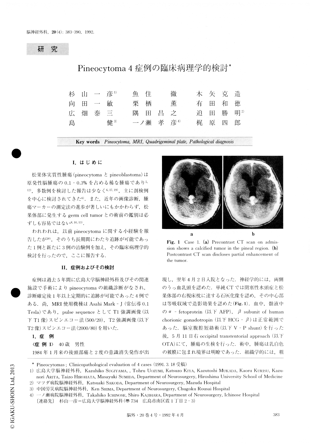
The subjects, 2 males and 2 females, ranged in age from 17 to 40. All the patients were clinically found to have the symptom of increased intracranial pressure on a monthly basis, but none of them were found to have dorsal midbrain dysfunction symptoms such as Pari-naucl's sign or Argyll Robertson pupil. Diagnostic imag-ing produced heterogenous pictures indicating calcifica-tions and cyst in 2 patients and homogenous pictures of the tumor parenchyma in the other 2 patients. Histolo-gically, the former cases were found to have many pineal-sand-like calcifications. Median sagittal MR im-ages demonstrated expansive growth of pineocytoma. Quadrigeminal plates which kept their shapes were observed in 2 patients. Craniotomy was performed in all cases, removing the tumor totally in 2 patients. Radiation therapy was given to 3 patients, resulting in complete remission, but radiosensitivity varied accord-ing to cases. During the follow-up period of 12 to 42 months, one patient died of peritonitis caused by shunt infection. No recurrence of the tumor was seen in any of the patients.
The incidence of pineocytoma was very low. Further evaluation of the tumor involving many cases is advis-able.

Copyright © 1992, Igaku-Shoin Ltd. All rights reserved.


