Japanese
English
- 有料閲覧
PPV購入案内
1,320円 (1,200円+税10%) こちらは、1097ページから1100ページの文献です。購入には医書.jp本会員登録が必要です。
会員登録完了後に改めて上記「購入サイトへ」を選択してください。
または「購入サイトへ」選択後に「購入ログイン」、「新規会員登録」の順に進んでください。
会員登録について詳しくはをご確認ください。
- Abstract 文献概要
- 1ページ目 Look Inside
I.はじめに
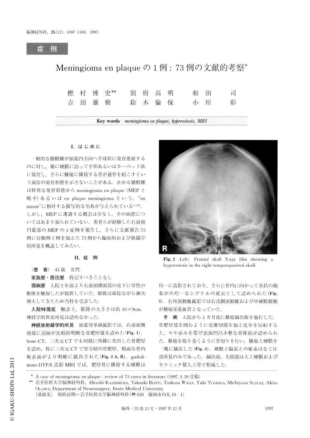
一般的な髄膜腫が頭蓋内方向へ半球状に発育進展するのに対し,稀に硬膜に沿って平坦あるいはカーペット状に発育し,さらに腫瘍に隣接する骨が過骨を起こすという通常の発育形態を示さないことがある.かかる髄膜腫は特異な発育形態からmeningioma en plaque(MEPと略す)あるいはen plaque meningiomaという,“en masse”に相対する描写的な名称が与えられている3,19).しかし,MEPに遭遇する機会は少なく,その病態についてはあまり知られていない.著者らが経験した右前頭円蓋部のMEPの1症例を報告し,さらに文献報告72例に自験例1例を加えた73例から臨床的および組織学的所見を概説してみたい.
The patient was a 41-year-old female who became aware of a protuberance in the right frontoparietal vault 2 years ago. Three dimensional CT showed a hyperostosis involving the diploe of the skull, and the rough surface of the inner table. In the gadoli-nium-DTPA enhanced MR images, an entity with a flame-like appearance invading the swollen skull rose from thick homogenously-enhanced dura. The lesion was surgically resected in a circle, and removed with the adherent dura. Histologically, this lesion exhibited a lobular pattern of meningothelial cells, and roughly in-vaded into the Haversian canals of the skull through the dura. Postoperatively, there was no neurological or physical deficit. In this paper, we reviewed clinical and pathological features of 73 meningioma en plaque cases including the present case.
Copyright © 1997, Igaku-Shoin Ltd. All rights reserved.