Japanese
English
- 有料閲覧
PPV購入案内
1,320円 (1,200円+税10%) こちらは、905ページから908ページの文献です。購入には医書.jp本会員登録が必要です。
会員登録完了後に改めて上記「購入サイトへ」を選択してください。
または「購入サイトへ」選択後に「購入ログイン」、「新規会員登録」の順に進んでください。
会員登録について詳しくはをご確認ください。
- Abstract 文献概要
- 1ページ目 Look Inside
I.はじめに
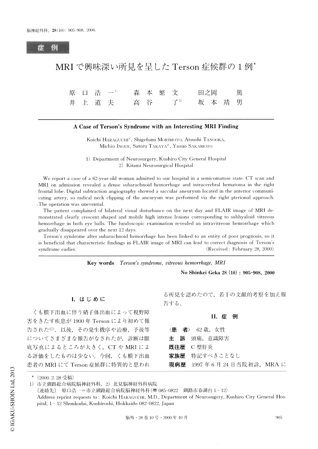
くも膜下出血に伴う硝子体出血によって視野障害をきたす疾患が1900年Tersonにより初めて報告された17).以後,その発生機序や治療,予後等についてさまざまな報告がなされたが,診断は眼底写真によるところが大きく,CTやMRIによる評価をしたものは少ない.今回,くも膜下出血患者のMRIにてTerson症候群に特異的と思われる所見を認めたので,若干の文献的考察を加え報告する.
We report a case of a 62-year-old woman admitted to our hospital in a semicomatose state. CT scan and MRI on admission revealed a dense subarachnoid hemorrhage and intracerebral hematoma in the right frontal lobe. Digital subtraction angiography showed a saccular aneurysm located in the anterior communi-cating artery, so radical neck clipping of the aneurysm was performed via the right pterional approach. The operation was unevential.
The patient complained of bilateral visual disturbance on the next day and FLAIR image of MRI de-monstrated clearly crescent shaped and mobile high intense lesions corresponding to subhyaloid vitreous hemorrhage in both eye balls. The fundoscopic examination revealed an intravitreous hemorrhage which gradually disappeared over the next 12 days.
Terson's syndrome after subarachnoid hemorrhage has been linked to an entity of poor prognosis, so it is beneficial that characteristic findings in FLAIR image of MRI can lead to correct diagnosis of Terson's syndrome earlier.
Copyright © 2000, Igaku-Shoin Ltd. All rights reserved.