Japanese
English
- 有料閲覧
PPV購入案内
1,320円 (1,200円+税10%) こちらは、615ページから621ページの文献です。購入には医書.jp本会員登録が必要です。
会員登録完了後に改めて上記「購入サイトへ」を選択してください。
または「購入サイトへ」選択後に「購入ログイン」、「新規会員登録」の順に進んでください。
会員登録について詳しくはをご確認ください。
- Abstract 文献概要
- 1ページ目 Look Inside
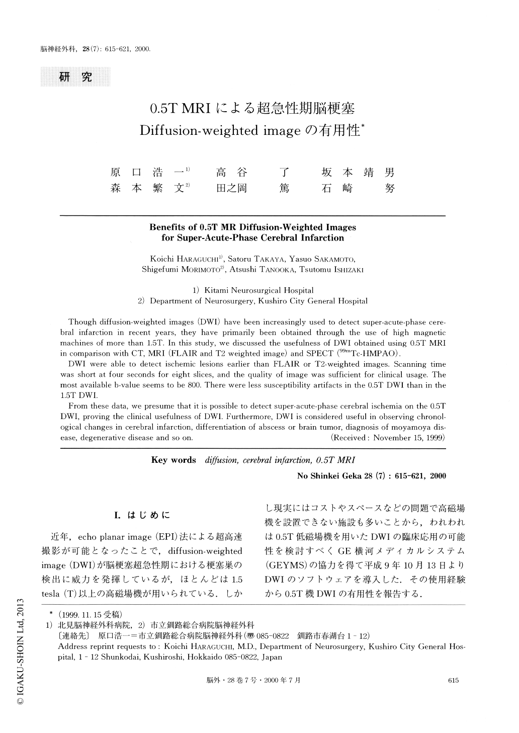
I.はじめに
近年,echo planar image(EPI)法による超高速撮影が可能となったことで,diffusion-weighted image(DWI)が脳梗塞超急性期における梗塞巣の検出に威力を発揮しているが,ほとんどは1.5tesla(T)以上の高磁場機が用いられている.しかし現実にはコストやスペースなどの問題で高磁場機を設置できない施設も多いことから,われわれは0.5T低磁場機を用いたDWIの臨床応用の可能性を検討すべくGE横河メディカルシステム(GEYMS)の協力を得て平成9年10月13日よりDWIのソフトウェアを導入した.その使用経験から0.5T機DWIの有用性を報告する.
Though diffusion-weighted images (DWI) have been increasingly used to detect super-acute-phase cere-bral infarction in recent years, they have primarily been obtained through the use of high magnetic machines of more than 1.5T. In this study, we discussed the usefulness of DWI obtained using 0.5T MRI in comparison with CT, MRI (FLAIR and T2 weighted image) and SPECT (99mTc-HMPAO).
DWI were able to detect ischemic lesions earlier than FLAIR or T2-weighted images. Scanning time was short at four seconds for eight slices, and the quality of image was sufficient for clinical usage. The most available b-value seems to be 800. There were less susceptibility artifacts in the 0.5T DWI than in the 1.5T DWI.
From these data, we presume that it is possible to detect super-acute-phase cerebral ischemia on the 0.5T DWI, proving the clinical usefulness of DWI. Furthermore, DWI is considered useful in observing chronol-ogical changes in cerebral infarction, differentiation of abscess or brain tumor, diagnosis of moyamoya dis-ease, degenerative disease and so on.
Copyright © 2000, Igaku-Shoin Ltd. All rights reserved.