Japanese
English
- 有料閲覧
PPV購入案内
1,320円 (1,200円+税10%) こちらは、401ページから406ページの文献です。購入には医書.jp本会員登録が必要です。
会員登録完了後に改めて上記「購入サイトへ」を選択してください。
または「購入サイトへ」選択後に「購入ログイン」、「新規会員登録」の順に進んでください。
会員登録について詳しくはをご確認ください。
- Abstract 文献概要
- 1ページ目 Look Inside
I.はじめに
3次元CT angiography(3D-CTA)は,従来とは異なる利点を有する画像診断法として,日常脳神経外科診療に欠かせないものとなっている.特に脳動脈瘤や頸部内頸動脈狭窄性病変の診断・治療に関しては,血管撮影やMRAと対比した詳細な検討がなされている1-4,8-12).
3D-CTAは造影剤の使用やX線被曝などの一定の侵襲性を有するが,従来の血管撮影と比べはるかに低侵襲かつ簡便である.現状では撮影範囲や造影剤投与量の制限,画像精度の問題から脳血管と頸部血管をそれぞれ単独で行う方法が一般的である.したがって頭蓋内動脈と頸動脈の情報を得たい場合,2回の検査施行が必要となるが,仮に1回で施行可能であれば,患者の検査リスク(煩雑さ,X線被曝,造影剤投与量)の軽減につながる.このような背景から,造影剤の単回投与にて頭頸部血管を同時に描出する方法(頭頸部血管単回造影CTA法)を考案し,1995年11月より臨床応用を開始して有用性を確認してきた.
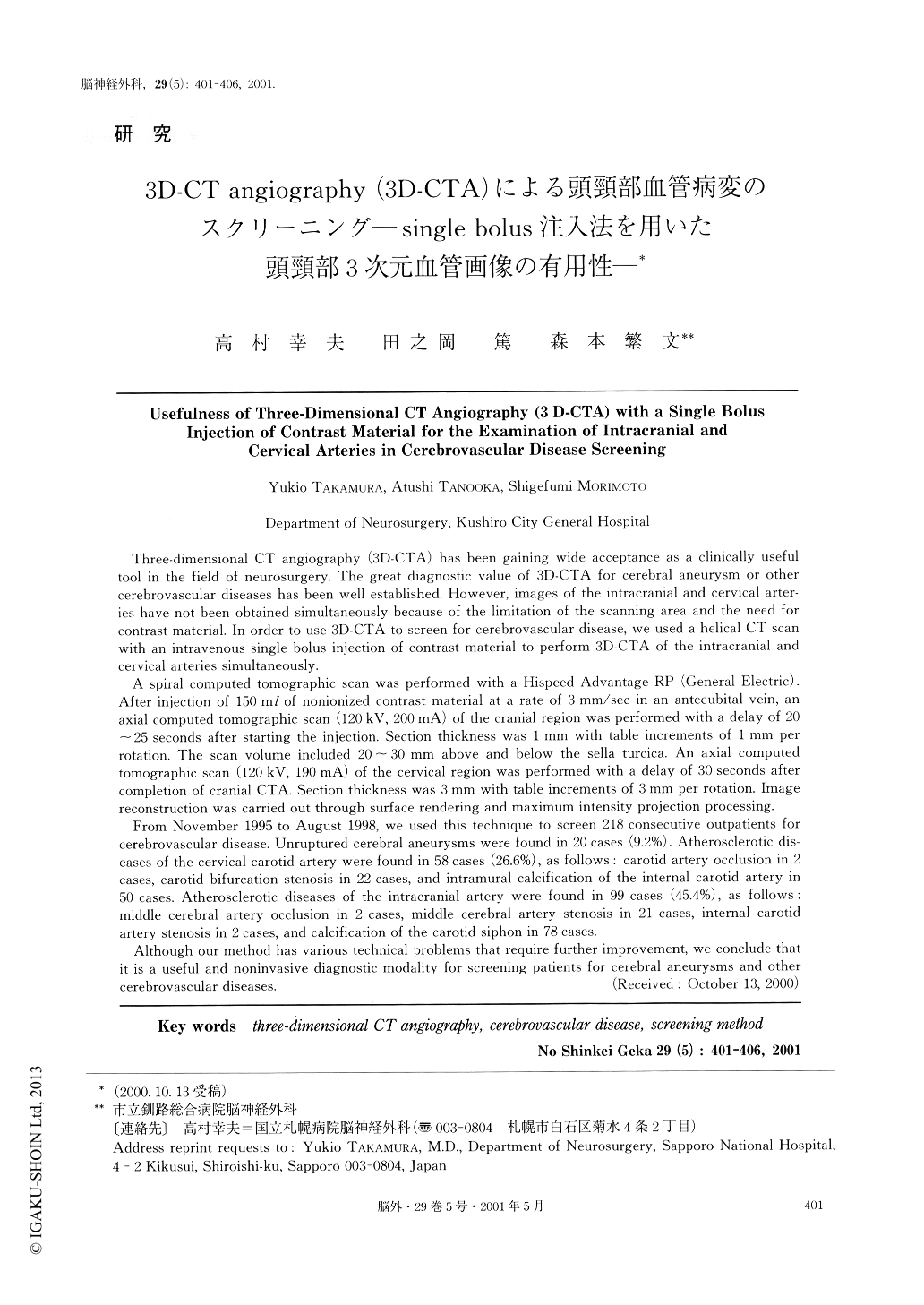
Three-dimensional CT angiography (3D-CTA) has been gaining wide acceptance as a clinically useful tool in the field of neurosurgery. The great diagnostic value of 3D-CTA for cerebral aneurysm or other cerebrovascular diseases has been well established. However, images of the intracranial and cervical arter- ies have not been obtained simultaneously because of the limitation of the scanning area and the need for contrast material. In order to use 3D-CTA to screenfor cerebrovascular disease, we used a helical CTscanwith an intravenous single bolus injection ofcontrast material to perform 3D-CTA of theintracranial andcervical arteries simultaneously.
A spiral computed tomographic scan was performedwith a Hispeed Advantage RP (General Electric).After injection of 150 ml of nonionized contrastmaterial at a rate of 3 mm/sec in an antecubitalvein, anaxial computed tomographic scan (120 kV, 200 mA) ofthe cranial region was performed with a delay of 20~25 seconds after starting the injection. Sectionthickness was 1 mm with table increments of 1 mm perrotation. The scan volume included 20~30 mm aboveand below the sella turcica. An axial computedtomographic scan (120 kV, 190 mA) of the cervicalregion was performed with a delay of 30 secondsaftercompletion of cranial CTA. Section thickness was 3mm with table increments of 3 mm per rotation. Imagereconstruction was carried out through surfacerendering and maximum intensity projectionprocessing.
From November 1995 to August 1998, we used thistechnique to screen 218 consecutive outpatients forcerebrovascular disease. Unruptured cerebralaneurysms were found in 20 cases (9.2%).Atherosclerotic dis-eases of the cervical carotid artery were found in58 cases (26.6%), as follows : carotid arteryocclusion in 2cases, carotid bifurcation stenosis in 22 cases, andintramural calcification of the internal carotidartery in50 cases. Atherosclerotic diseases of theintracranial artery were found in 99 cases (45.4%),as follows :middle cerebral artery occlusion in 2 cases, middlecerebral artery stenosis in 21 cases, internalcarotid artery stenosis in 2 cases, and calcification of thecarotid siphon in 78 cases. Although our method has various technical problemsthat require further improvement, we conclude thatit is a useful and noninvasive diagnostic modalityfor screening patients for cerebral aneurysms andothercerebrovascular diseases.
Copyright © 2001, Igaku-Shoin Ltd. All rights reserved.