Japanese
English
- 有料閲覧
PPV購入案内
1,320円 (1,200円+税10%) こちらは、543ページから547ページの文献です。購入には医書.jp本会員登録が必要です。
会員登録完了後に改めて上記「購入サイトへ」を選択してください。
または「購入サイトへ」選択後に「購入ログイン」、「新規会員登録」の順に進んでください。
会員登録について詳しくはをご確認ください。
- Abstract 文献概要
- 1ページ目 Look Inside
I.はじめに
頭蓋骨骨髄炎は以前には,副鼻腔炎に続発したり,他の臓器より血行性に感染する報告例が多く見られた7)が,近年,抗生物質の発達により,比較的稀な疾患となっている.また,外傷を契機に発症する場合には,開放性頭蓋骨骨折後や頭蓋底骨折症例に起こり,発生機序は明瞭である.今回,われわれは頭部外傷後約30年もの長い経過を示した頭蓋骨骨髄炎症例を経験したので報告する.
We report a case of cranial osteomyelitis after head injury with exaggeration and remission for 30 years.
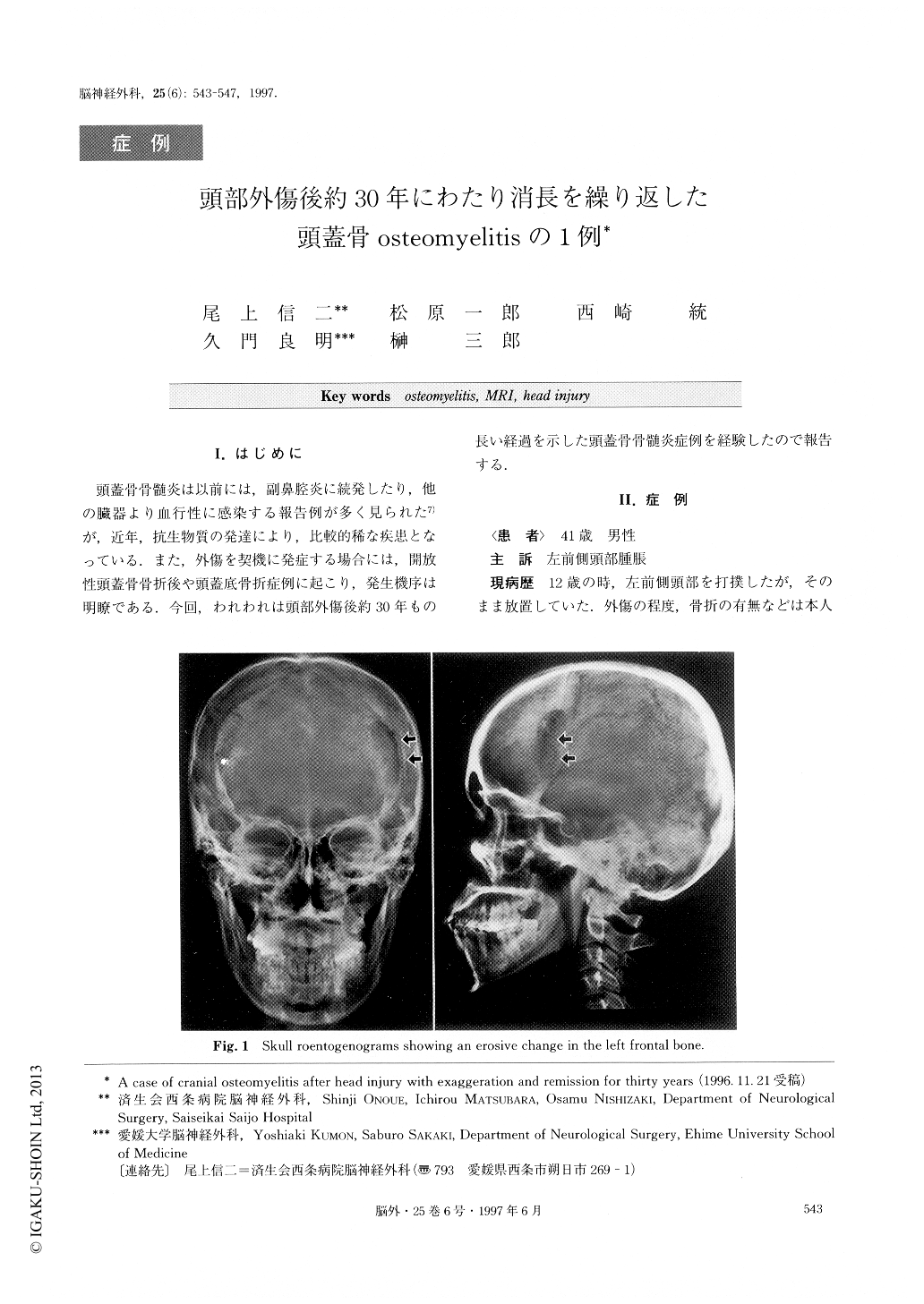
A 41-year-old man was referred to our hospital with redness, swelling and pain in the left frontal region. He had noted a hard mass in the same region after an in-jury at the age of 12 years, and local heat and redness again in the same region which had disappeared with-out definite treatment at the age of 20 years. Presently, skull roentogenogram showed an erosive lesion in the left frontal bone. Plain CT revealed bone defect, epidu-ral mass lesion, calcification of the dura mater and thickness of the extracranial soft tissue in the left fron-tal region. T1-weighted MR images showed an isoin-tense lesion and T2-weighted MR images showed a high intensity lesion in the inner table and a diploic layer in the left frontal bone.
99mTc MDP bone scinnti-gram showed abnormal uptake in the same region. Operation performed through a left frontotemporal craniotomy revealed a degeneration of temporal muscle and frontal bone, and granulation tissue and pus in the subcutaneous and epidural spaces. These were removed together with the calcified dura mater. The subdural space was intact. Dural plasty was performed using fas-cia lata, but the craniotomied bone was replaced. Histo-logical examination revealed typical findings of osteomyelitis. Postoperative course was uneventful, and he was discharged without neurological deficit. Cranio-plasty was performed 8 months after the first surgery. The mechanism by which osteomyelitis continued over a long course of time was suspected to be the formation of a focus of recurrent inflammation due to the head injury 30 years earlier.
Copyright © 1997, Igaku-Shoin Ltd. All rights reserved.