Japanese
English
- 有料閲覧
- Abstract 文献概要
- 1ページ目 Look Inside
I.はじめに
脳動静脈奇形(AVM)は,発症形式が出血か非出血かで以後の経過に大きな違いがある.出血例の再出血率は30-45%であるのに対し,非出血例の出血率は5-10%程度とされている3,7,12).一方,AVMの自然消失は2—3%に起こると報告されているが,AVMが自然消失する機序は必ずしも明らかでない1-11)今回われわれは,痙攣発作で発症し経過観察中に脳血管写上主幹動脈の閉塞とともにAVMの自然消失を認めた1例を経験したので自験例のAVMの自然消失の機序について若干の文献的考察を加え報告する.
A rare case of spontaneous regression of cerebral arterio-venous malformation (AVM) is reported.
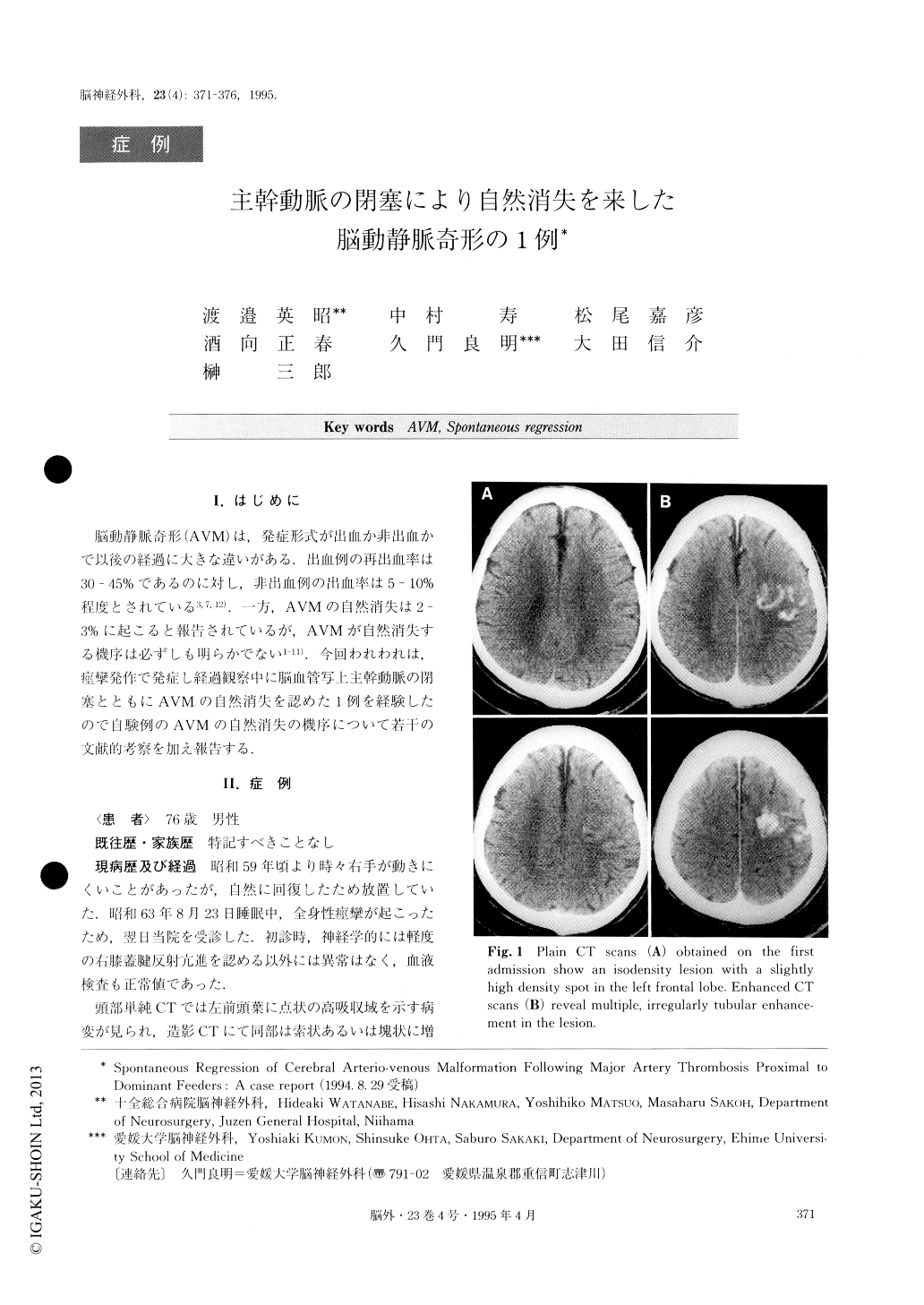
A 76-year-old male was admitted to Juzen General Hospital due to generalized convulsion on August 24, 1987. On admission, results of physical and neurological examinations were normal. Plain CT scans showed an iso-density lesion with a slightly high density spot in the left frontal lobe, and enhanced CT scans showed multiple, irregularly tubular enhancements in the lesion.Left carotid angiogram (CAG) demonstrated an arte-riovenous malformation (AVM) in the left frontal lobe, which was fed by the left ACA and MCA and drained by the ascending cerebral vein and superficial Sylvian vein. There was also an anterior communicating artery aneurysm. At the patient's request, he was treated con-servatively and was discharged without neurological deficit. Though he continued to take anticonvulsants thereafter, he felt weakness or numbness of the right extremities once a year. Five years after the first admis-sion, he developed sudden onset of right hemiparesis, aphasia and consciousness disturbance. On admission, his platelet aggregation function was elevated. At this time, enhanced CT scans did not show any enhanced area in the left frontal lobe where AVM had been found previously. T2-weighted magnetic resonance im-age showed a mixed intensity area without any flow void phenomenon suggesting thrombosis of the nidus. Left CAG demonstrated occulsion of the A1-A2 junc-tion of the anterior cerebral artery and disappearance of the AVM. He was treated conservatively again, and was discharged without neurological deficit. The mechanism of the regression of AVM in this case seemed to be thrombosis of the nidus following major artery thrombosis proximal to main feeders. This was caused by arteriosclerotic change, elevated platelet aggregation, and local hemodynamic change due to an aneurysm of the anterior communicating artery.

Copyright © 1995, Igaku-Shoin Ltd. All rights reserved.


