Japanese
English
- 有料閲覧
PPV購入案内
1,320円 (1,200円+税10%) こちらは、685ページから691ページの文献です。購入には医書.jp本会員登録が必要です。
会員登録完了後に改めて上記「購入サイトへ」を選択してください。
または「購入サイトへ」選択後に「購入ログイン」、「新規会員登録」の順に進んでください。
会員登録について詳しくはをご確認ください。
- Abstract 文献概要
- 1ページ目 Look Inside
I.はじめに
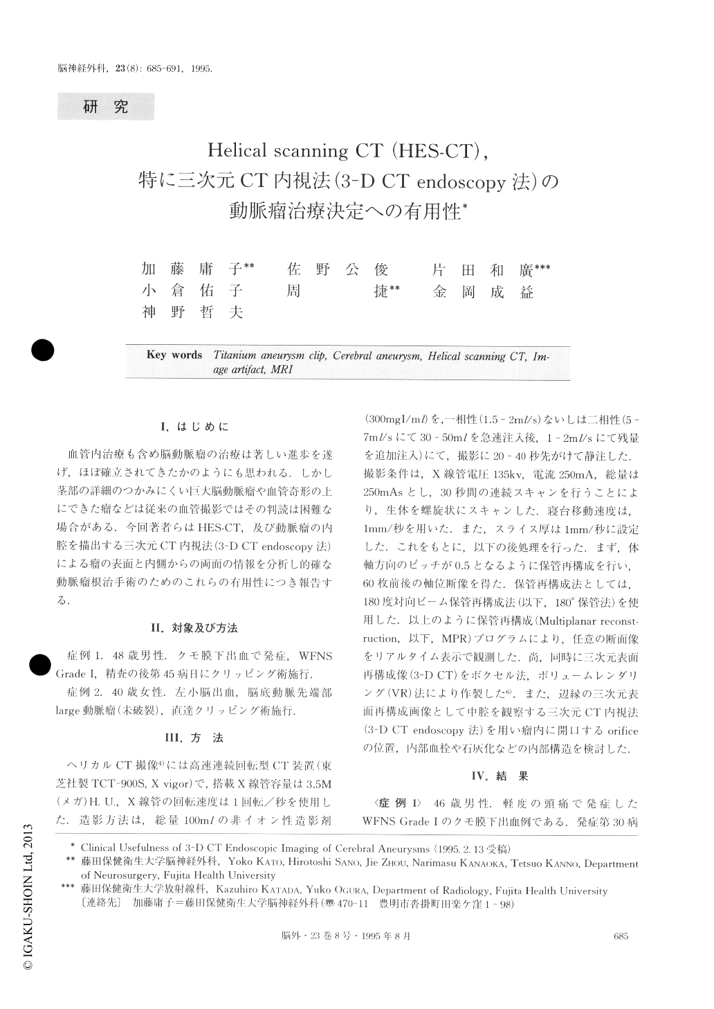
血管内治療も含め脳動脈瘤の治療は著しい進歩を遂げ,ほぼ確立されてきたかのようにも思われる.しかし茎部の詳細のつかみにくい巨大脳動脈瘤や血管奇形の上にできた瘤などは従来の血管撮影ではその判読は困難な場合がある.今回著者らはHES-CT,及び動脈瘤の内腔を描出する三次元CT内視法(3—DCT endoscopy法)による瘤の表面と内側からの両面の情報を分析し的確な動脈瘤根治手術のためのこれらの有用性につき報告する,
Usefulness of endoscopic imaging of cerebral aneu-rysms is presented. 3D-luminal images were obtained using a new processing technique which extracts CT numbers in the boundary region between the vessel wall and contrast media filling in the vascular lumen. Clinical application of this technique to complicated large cerebral aneurysms showed that, with this 3D-CT endoscopic images and MRA, anatomical details of cerebral aneurysms such as the orifice of the aneurysm, intraluminal thrombus, and calcification of the wall could be clearly demonstrated. We operated on two large, complicated aneurysms after obtaining 3D-CT endoscopy images of the aneurysms. Such information was found to be very useful when operating on difficult and complicated cerebral aneurysms.
Copyright © 1995, Igaku-Shoin Ltd. All rights reserved.