Japanese
English
- 有料閲覧
PPV購入案内
1,320円 (1,200円+税10%) こちらは、1083ページから1087ページの文献です。購入には医書.jp本会員登録が必要です。
会員登録完了後に改めて上記「購入サイトへ」を選択してください。
または「購入サイトへ」選択後に「購入ログイン」、「新規会員登録」の順に進んでください。
会員登録について詳しくはをご確認ください。
- Abstract 文献概要
- 1ページ目 Look Inside
Ⅰ.はじめに
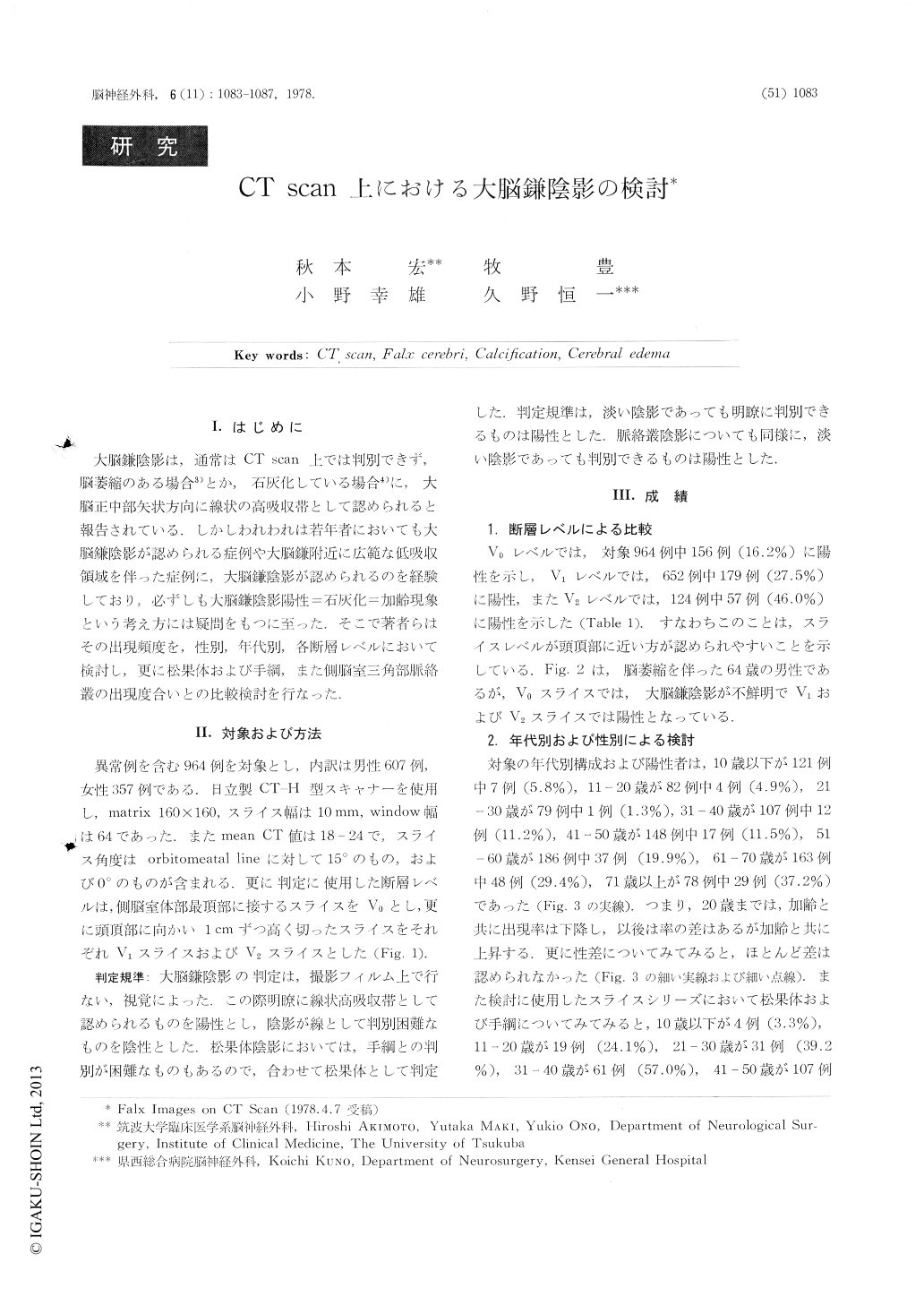
大脳鎌陰影は,通常はCT scan上では判別できず,脳萎縮のある場合3)とか,石灰化している場合4)に,大脳正中部矢状方向に線状の高吸収帯として認められると報告されている.しかしわれわれは若年者においても大脳鎌陰影が認められる症例や大脳鎌附近に広範な低吸収領域を伴った症例に,大脳鎌陰影が認められるのを経験しており,必ずしも大脳鎌陰影陽性=石灰化=加齢現象という考え方には疑問をもつに至った.そこで著者らはその出現頻度を,性別,年代別,各断層レベルにおいて検討し,更に松果体および手綱,また側脳室三角部脈絡叢の出現度合いとの比較検討を行なった.
It has been reported that falx images usually are not visualized on CT scan except in the case with brain atrophy of falx calcification. But we have often experi-enced the falx images on CT scan in the case of normal child, and we have an idea that flax images do not always mean a calcification of the falx.
Evaluation of falx images on CT scan was done in 964 normal or abnormal cases in relation to different CT slice level, age, sex, pineal body and habenula and choroid plexus.
Copyright © 1978, Igaku-Shoin Ltd. All rights reserved.