Japanese
English
- 有料閲覧
PPV購入案内
1,320円 (1,200円+税10%) こちらは、905ページから913ページの文献です。購入には医書.jp本会員登録が必要です。
会員登録完了後に改めて上記「購入サイトへ」を選択してください。
または「購入サイトへ」選択後に「購入ログイン」、「新規会員登録」の順に進んでください。
会員登録について詳しくはをご確認ください。
- Abstract 文献概要
- 1ページ目 Look Inside
I.はじめに
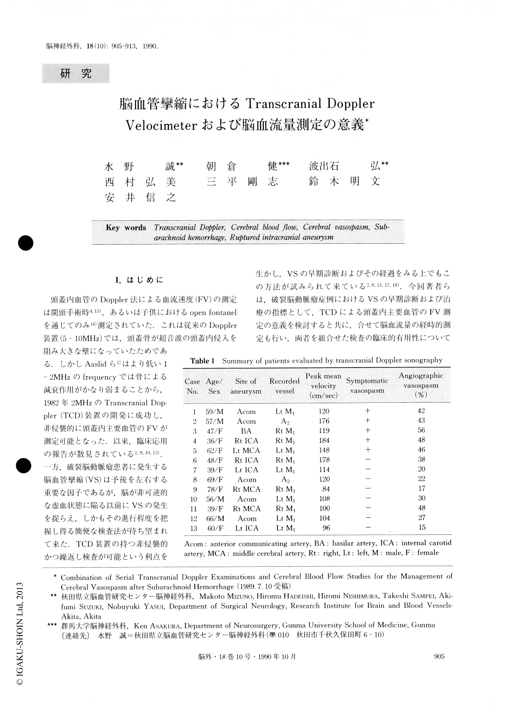
頭蓋内血管のDoppler法による血流速度(FV)の測定は開頭手術時4,15),あるいは子供におけるopen fontanelを通じてのみ14)測定されていた.これは従来のDoppler装置(5-10MHz)では,頭蓋骨が超音波の頭蓋内侵入を阻み大きな壁になっていたためである.しかしAaslidら1)はより低い1−2MHzのfrequencyでは骨による減衰作用がかなり弱まることから,1982年2MHzのTranscranial Dop—pler(TCD)装置の開発に成功し,非侵襲的に頭蓋内主要血管のFVが測定可能となった.以来,臨床応用の報告が散見されている2,9,10,13).一方,破裂脳動脈瘤患者に発生する脳血管攣縮(VS)は予後を左右する重要な因子であるが,脳が非可逆的な虚血状態に陥る以前にVSの発生を捉らえ,しかもその進行程度を把握し得る簡便な検査法が待ち望まれて来た.
In 13 patients who had ruptured intracranial aneurysms, serial transcranial Doppler (TCD) and cere-bral blood flow (CBF) examinations were performed in order to evaluate the degree of cerebral vasospasm. All patients showed some extent of vasospasm on angiography, which was performed between Day 7 and 10. The flow velocities of either the middle cerebral arteries or the anterior cerebral arteries, measured by TCD, began to increase on post hemorrhage Day 5, and maximum flow velocities were recorded between Day 9 and 13, with normalization occurring within the following 2 weeks.
Copyright © 1990, Igaku-Shoin Ltd. All rights reserved.