Japanese
English
- 有料閲覧
PPV購入案内
1,320円 (1,200円+税10%) こちらは、205ページから210ページの文献です。購入には医書.jp本会員登録が必要です。
会員登録完了後に改めて上記「購入サイトへ」を選択してください。
または「購入サイトへ」選択後に「購入ログイン」、「新規会員登録」の順に進んでください。
会員登録について詳しくはをご確認ください。
- Abstract 文献概要
- 1ページ目 Look Inside
- 参考文献 Reference
Ⅰ.はじめに
副鼻腔炎に合併する頭蓋内感染症,特に硬膜下蓄膿(膿瘍)は,画像診断の向上と抗菌薬の発達した近年においても,重篤な後遺症を生じ,致死率も高い.そのため,迅速な診断と的確な治療が必要な脳神経外科領域の重要な救急疾患である4,6).
今回われわれは,右歯性上顎洞炎から左硬膜下蓄膿と左中大脳動脈の続発性動脈炎による脳虚血を生じた1例を経験したので,文献的考察を加え報告する.
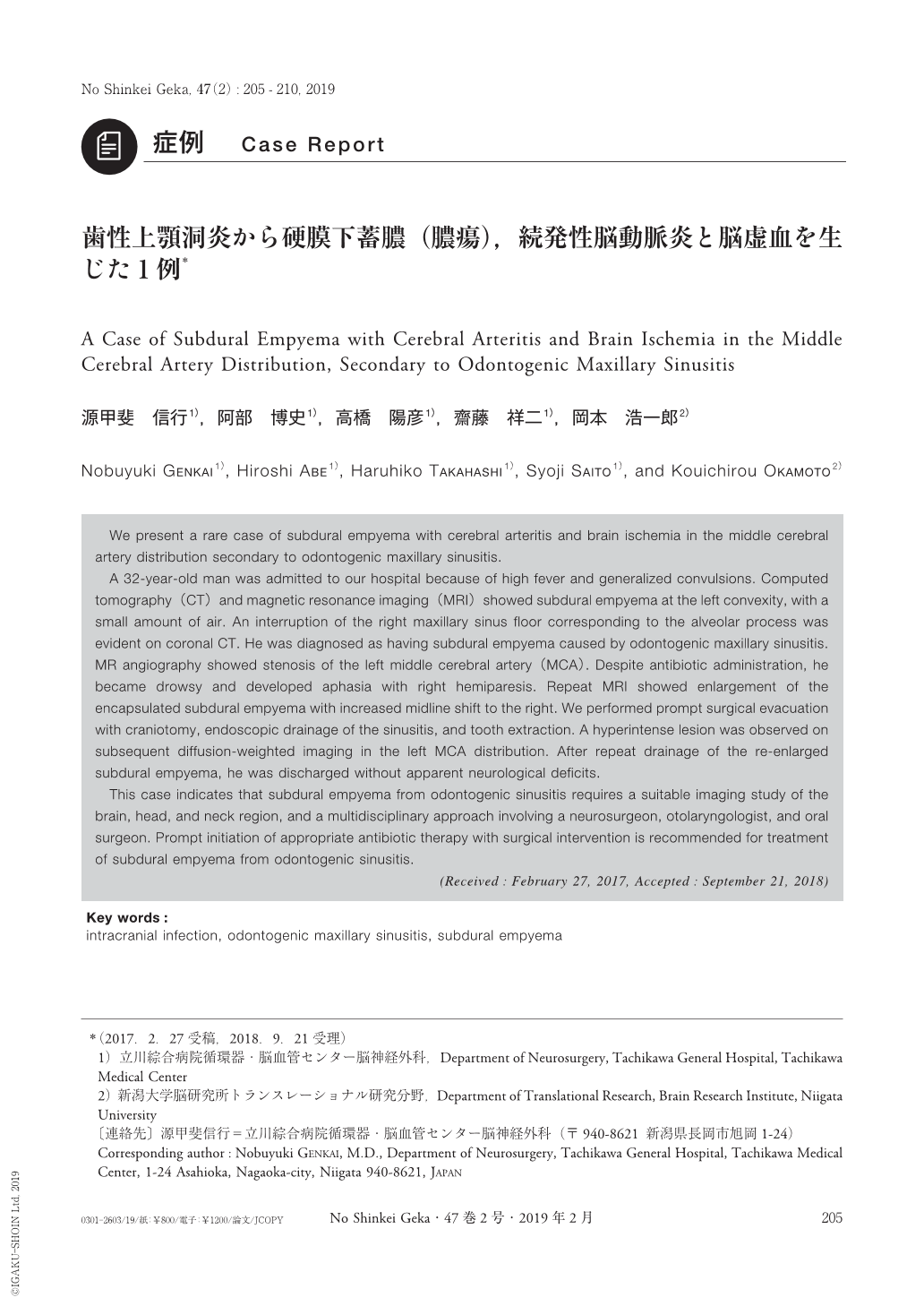
We present a rare case of subdural empyema with cerebral arteritis and brain ischemia in the middle cerebral artery distribution secondary to odontogenic maxillary sinusitis.
A 32-year-old man was admitted to our hospital because of high fever and generalized convulsions. Computed tomography(CT)and magnetic resonance imaging(MRI)showed subdural empyema at the left convexity, with a small amount of air. An interruption of the right maxillary sinus floor corresponding to the alveolar process was evident on coronal CT. He was diagnosed as having subdural empyema caused by odontogenic maxillary sinusitis. MR angiography showed stenosis of the left middle cerebral artery(MCA). Despite antibiotic administration, he became drowsy and developed aphasia with right hemiparesis. Repeat MRI showed enlargement of the encapsulated subdural empyema with increased midline shift to the right. We performed prompt surgical evacuation with craniotomy, endoscopic drainage of the sinusitis, and tooth extraction. A hyperintense lesion was observed on subsequent diffusion-weighted imaging in the left MCA distribution. After repeat drainage of the re-enlarged subdural empyema, he was discharged without apparent neurological deficits.
This case indicates that subdural empyema from odontogenic sinusitis requires a suitable imaging study of the brain, head, and neck region, and a multidisciplinary approach involving a neurosurgeon, otolaryngologist, and oral surgeon. Prompt initiation of appropriate antibiotic therapy with surgical intervention is recommended for treatment of subdural empyema from odontogenic sinusitis.
Copyright © 2019, Igaku-Shoin Ltd. All rights reserved.