Japanese
English
- 有料閲覧
- Abstract 文献概要
- 1ページ目 Look Inside
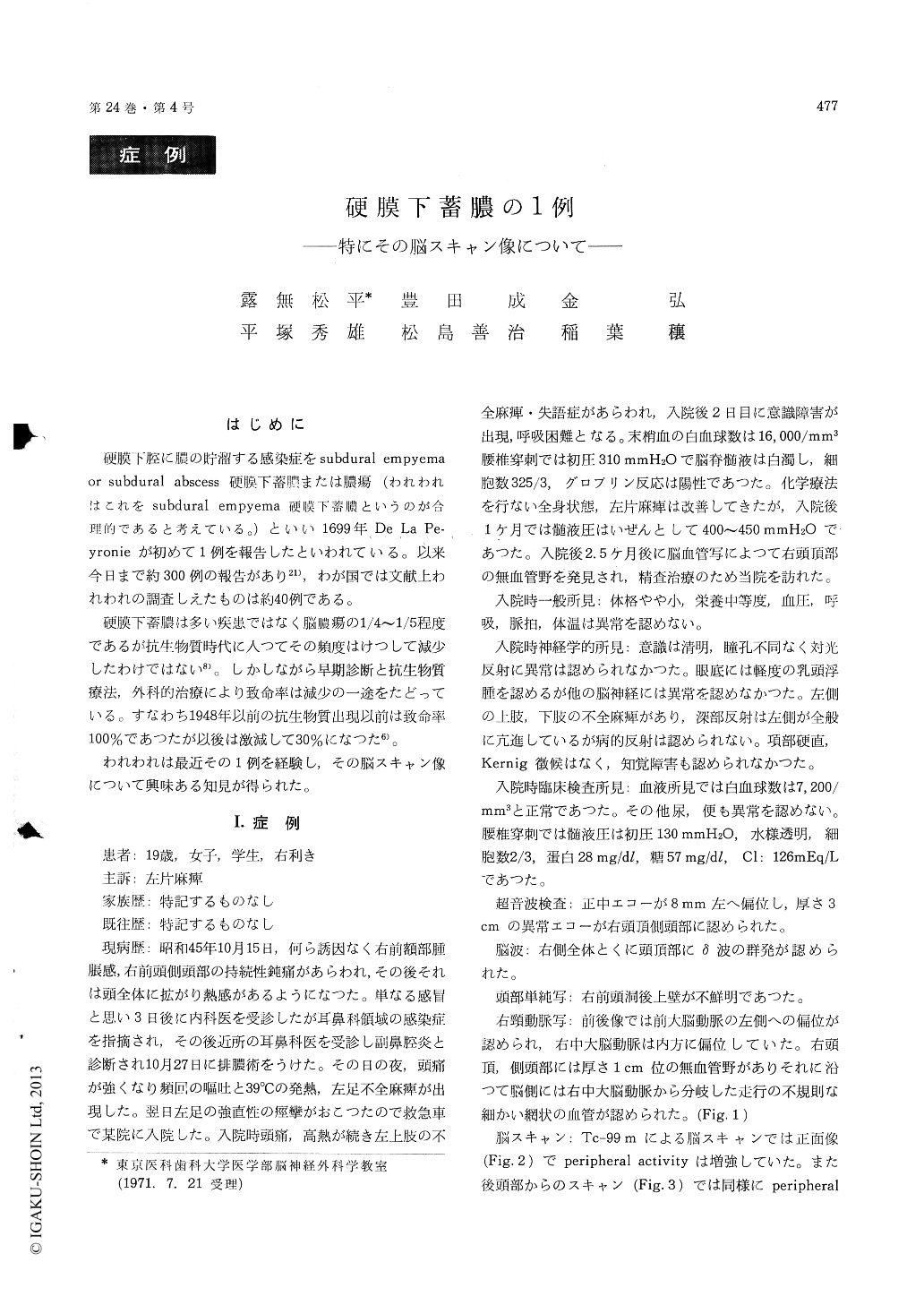
はじめに
硬膜下腔に膿の貯溜する感染症をsubdural empyemaor subdural abscess硬膜下蓄膿または膿瘍(われわれはこれをsubdural empyema硬膜下蓄膿というのが合理的であると考えている。)といい1699年、De La Pe—yronieが初めて1例を報告したといわれている。以来今日まで約300例の報告があり21),わが国では文献上われわれの調査しえたものは約40例である。
硬膜下蓄膿は多い疾患ではなく脳膿瘍の1/4〜1/5程度であるが抗生物質時代に入つてその頻度はけつして減少したわけではない8)。しかしながら早期診断と抗生物質療法,外科的治療により致命率は減少の一途をたどっている。すなわち1948年以前の抗生物質出現以前は致命率100%であつたが以後は激減して30%になつた6)。
Subdural empyema is rather infrequent intra-cranial suppuration. About 40 cases have been reported ever since in Japan. The literature on scan appearance of subdural empyema has been extremely rare. Our purpose is to briefly review the scan findings in subdural empyema and to re-port a case with a triad of scan findings.
This 19 year old right-handed female complained of dull pain on the right frontal and temporal area on Oct. 15, 1970. It was followed by elevated body temperature, vomiting, left-sided hemiparesis and tonic convulsion. After the admission to a localhospital, motor aphasia, disturbance of consciousnessand dyspnea developed. Systemic antibiotic therapyresulted in the disappearance of these symptoms. The right-sided carotid angiograihy revealed pro-nounced avascular area on the right parietal region.After being transferred to our hospital, two burrholes were made on the right parietal area and100 ml of thick yellow pus was evacuated. Cul-tures were made for sensitivity study but werenegative. Subdural space was lavaged with copiousamounts of sterile saline and then with cephalo-ridine solution. Two drains were removed threedays after the operation. She was discharged withslight hemiparesis on the left, 4 weeks after theoperation.
Scan findings on this patient are as follows.
1) increased peripheral activity
2) so-called "doughnut sign" was positive.
3) The posterior view shows the crescent-shapedactivity, 2×5cm in size in the midline. Themechanism of this pattern, we are inferring, is asfollows. In this case pus extended from frontal tooccipital along the falx forming interhemisphericsubdural empyema, and then went farther to theopposite side along the contiguous epitentorialspace. On the other hand the extension to theipsilateral side was prevented because of elevatedintracranial pressure on that side.
In conclusion, brain scan is the most usefuladjunct in diagnosing subdural empyema, for whichfour directional brain scans are essential.
Copyright © 1972, Igaku-Shoin Ltd. All rights reserved.