Japanese
English
- 有料閲覧
PPV購入案内
1,320円 (1,200円+税10%) こちらは、509ページから514ページの文献です。購入には医書.jp本会員登録が必要です。
会員登録完了後に改めて上記「購入サイトへ」を選択してください。
または「購入サイトへ」選択後に「購入ログイン」、「新規会員登録」の順に進んでください。
会員登録について詳しくはをご確認ください。
- Abstract 文献概要
- 1ページ目 Look Inside
- 参考文献 Reference
Ⅰ.はじめに
類上皮腫は脳腫瘍の中で0.2〜1.8%を占める良性腫瘍である3).組織学的には角化を伴う重層扁平上皮で被覆された囊胞であり,皮膚付属器は伴わない.好発部位としては小脳橋角部,中頭蓋窩,トルコ鞍近傍,脊髄,頭蓋骨板間層などがあり,稀に悪性転化して扁平上皮癌が発生する3).今回われわれは,悪性転化し,髄腔内播種を来したと思われる類上皮腫の稀な1例を経験したため,文献的考察を含めて報告する.
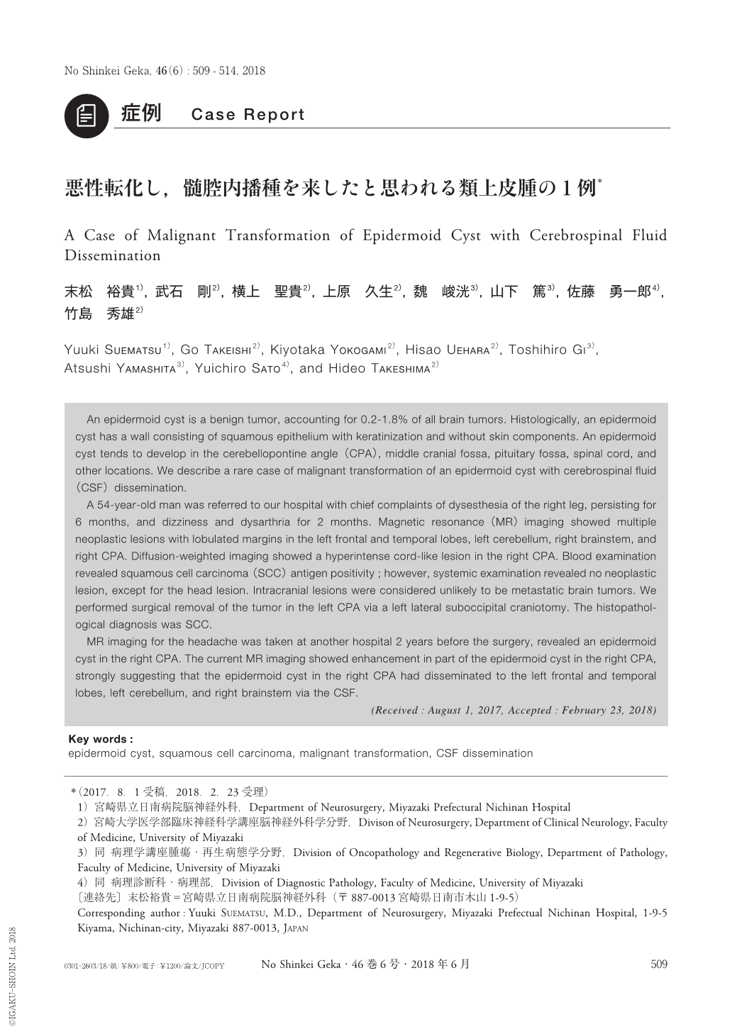
An epidermoid cyst is a benign tumor, accounting for 0.2-1.8% of all brain tumors. Histologically, an epidermoid cyst has a wall consisting of squamous epithelium with keratinization and without skin components. An epidermoid cyst tends to develop in the cerebellopontine angle(CPA), middle cranial fossa, pituitary fossa, spinal cord, and other locations. We describe a rare case of malignant transformation of an epidermoid cyst with cerebrospinal fluid(CSF)dissemination.
A 54-year-old man was referred to our hospital with chief complaints of dysesthesia of the right leg, persisting for 6 months, and dizziness and dysarthria for 2 months. Magnetic resonance(MR)imaging showed multiple neoplastic lesions with lobulated margins in the left frontal and temporal lobes, left cerebellum, right brainstem, and right CPA. Diffusion-weighted imaging showed a hyperintense cord-like lesion in the right CPA. Blood examination revealed squamous cell carcinoma(SCC)antigen positivity;however, systemic examination revealed no neoplastic lesion, except for the head lesion. Intracranial lesions were considered unlikely to be metastatic brain tumors. We performed surgical removal of the tumor in the left CPA via a left lateral suboccipital craniotomy. The histopathological diagnosis was SCC.
MR imaging for the headache was taken at another hospital 2 years before the surgery, revealed an epidermoid cyst in the right CPA. The current MR imaging showed enhancement in part of the epidermoid cyst in the right CPA, strongly suggesting that the epidermoid cyst in the right CPA had disseminated to the left frontal and temporal lobes, left cerebellum, and right brainstem via the CSF.
Copyright © 2018, Igaku-Shoin Ltd. All rights reserved.