Japanese
English
- 有料閲覧
PPV購入案内
1,320円 (1,200円+税10%) こちらは、19ページから28ページの文献です。購入には医書.jp本会員登録が必要です。
会員登録完了後に改めて上記「購入サイトへ」を選択してください。
または「購入サイトへ」選択後に「購入ログイン」、「新規会員登録」の順に進んでください。
会員登録について詳しくはをご確認ください。
- Abstract 文献概要
- 1ページ目 Look Inside
- 参考文献 Reference
Ⅰ.はじめに
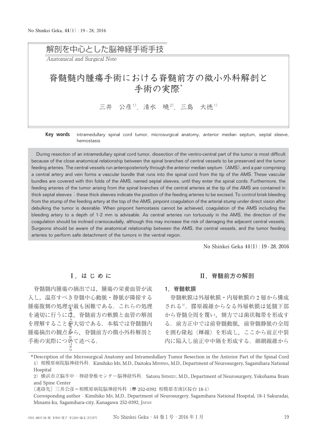
脊髄髄内腫瘍の摘出では,腫瘍の栄養血管が流入し,温存すべき脊髄中心動脈・静脈が隣接する腫瘍腹側の処理が最も困難である.これらの処理を適切に行うには,脊髄前方の軟膜と血管の解剖を理解することが大切である.本稿では脊髄髄内腫瘍摘出の観点から,脊髄前方の微小外科解剖と手術の実際について述べる.
During resection of an intramedullary spinal cord tumor, dissection of the ventro-central part of the tumor is most difficult because of the close anatomical relationship between the spinal branches of central vessels to be preserved and the tumor feeding arteries. The central vessels run anteroposteriorly through the anterior median septum(AMS), and a pair comprising a central artery and vein forms a vascular bundle that runs into the spinal cord from the tip of the AMS. These vascular bundles are covered with thin folds of the AMS, named septal sleeves, until they enter the spinal cords. Furthermore, the feeding arteries of the tumor arising from the spinal branches of the central arteries at the tip of the AMS are contained in thick septal sleeves;these thick sleeves indicate the position of the feeding arteries to be excised. To control brisk bleeding from the stump of the feeding artery at the top of the AMS, pinpoint coagulation of the arterial stump under direct vision after debulking the tumor is desirable. When pinpoint hemostasis cannot be achieved, coagulation of the AMS including the bleeding artery to a depth of 1-2 mm is advisable. As central arteries run tortuously in the AMS, the direction of the coagulation should be inclined craniocaudally, although this may increase the risk of damaging the adjacent central vessels. Surgeons should be aware of the anatomical relationship between the AMS, the central vessels, and the tumor feeding arteries to perform safe detachment of the tumors in the ventral region.
Copyright © 2016, Igaku-Shoin Ltd. All rights reserved.