Japanese
English
- 有料閲覧
PPV購入案内
1,320円 (1,200円+税10%) こちらは、87ページから91ページの文献です。購入には医書.jp本会員登録が必要です。
会員登録完了後に改めて上記「購入サイトへ」を選択してください。
または「購入サイトへ」選択後に「購入ログイン」、「新規会員登録」の順に進んでください。
会員登録について詳しくはをご確認ください。
- Abstract 文献概要
- 1ページ目 Look Inside
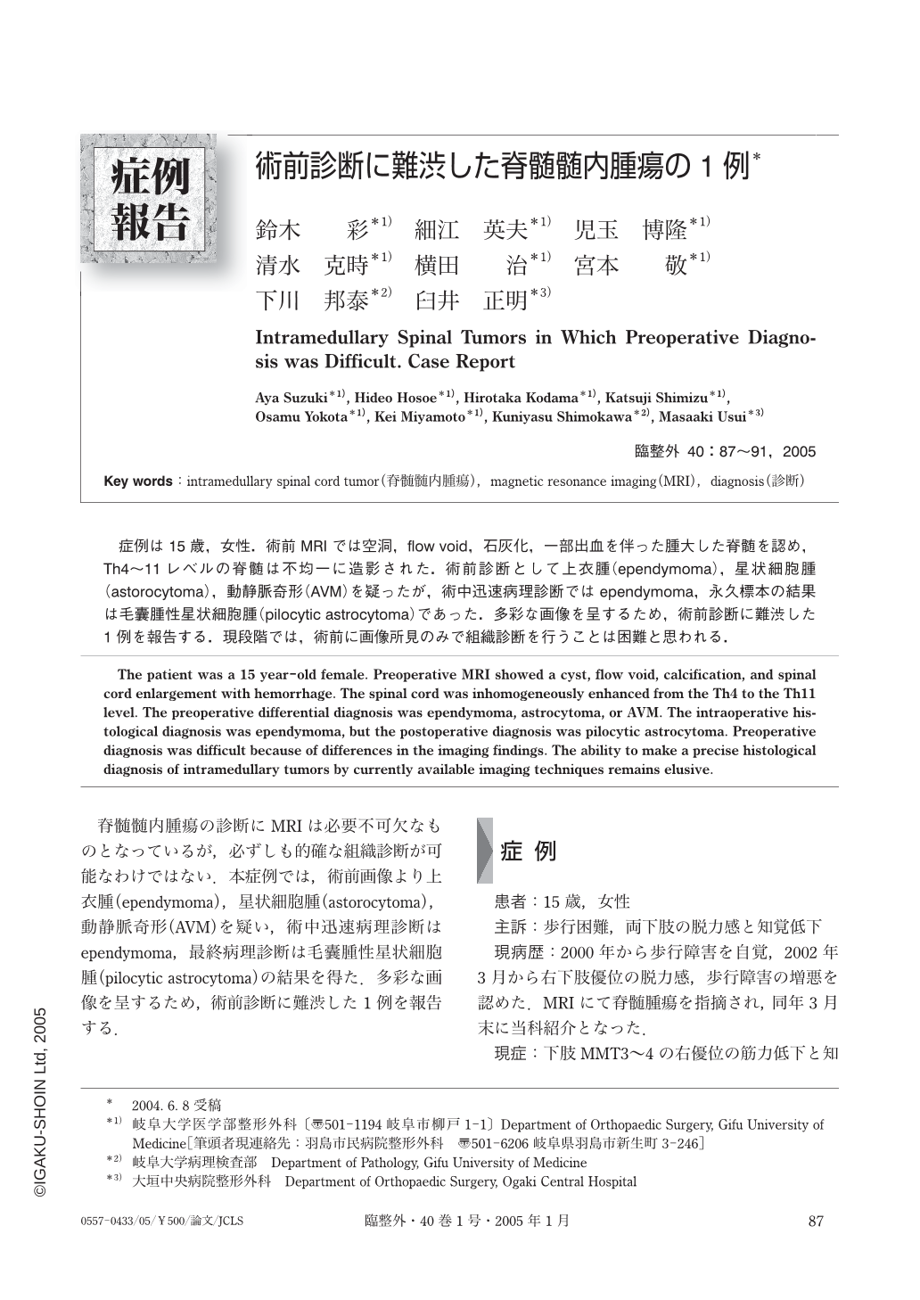
症例は15歳,女性.術前MRIでは空洞,flow void,石灰化,一部出血を伴った腫大した脊髄を認め,Th4~11レベルの脊髄は不均一に造影された.術前診断として上衣腫(ependymoma),星状細胞腫(astorocytoma),動静脈奇形(AVM)を疑ったが,術中迅速病理診断ではependymoma,永久標本の結果は毛嚢腫性星状細胞腫(pilocytic astrocytoma)であった.多彩な画像を呈するため,術前診断に難渋した1例を報告する.現段階では,術前に画像所見のみで組織診断を行うことは困難と思われる.
The patient was a 15 year-old female. Preoperative MRI showed a cyst, flow void, calcification, and spinal cord enlargement with hemorrhage. The spinal cord was inhomogeneously enhanced from the Th4 to the Th11 level. The preoperative differential diagnosis was ependymoma, astrocytoma, or AVM. The intraoperative histological diagnosis was ependymoma, but the postoperative diagnosis was pilocytic astrocytoma. Preoperative diagnosis was difficult because of differences in the imaging findings. The ability to make a precise histological diagnosis of intramedullary tumors by currently available imaging techniques remains elusive.
Copyright © 2005, Igaku-Shoin Ltd. All rights reserved.